This site uses cookies to improve your experience. To help us insure we adhere to various privacy regulations, please select your country/region of residence. If you do not select a country, we will assume you are from the United States. Select your Cookie Settings or view our Privacy Policy and Terms of Use.
Cookie Settings
Cookies and similar technologies are used on this website for proper function of the website, for tracking performance analytics and for marketing purposes. We and some of our third-party providers may use cookie data for various purposes. Please review the cookie settings below and choose your preference.
Used for the proper function of the website
Used for monitoring website traffic and interactions
Cookie Settings
Cookies and similar technologies are used on this website for proper function of the website, for tracking performance analytics and for marketing purposes. We and some of our third-party providers may use cookie data for various purposes. Please review the cookie settings below and choose your preference.
Strictly Necessary: Used for the proper function of the website
Performance/Analytics: Used for monitoring website traffic and interactions
Then I always look to see if the initial deflection of the QRS has a lot of voltage change per change in time (seen in tachycardias that are initiated from above the ventricle because the propagate through fast conducting purkinje fiber. Tachycardia exaggerates ST Elevation in LBBB and Paced rhythm 5. Pacemaker mediated tachycardia!
Shortly after isoprenalin infusion was initiated, there were short runs of ventricular tachycardia. Figure-3: Diagnostic considerations for a patient who presents in AV block ( adapted from Mangi et al — StatPearls, 2021 ). She was started on isoprenalin (isoproterenol). The above ECG initially shows AV block.
We would like to comment on the publication “Postural orthostatic tachycardia syndrome after COVID-19 vaccination [1].” This study evaluated patients who received the mRNA COVID-19 vaccine and then developed new or worsening symptoms of Postural Orthostatic Tachycardia Syndrome (POTS).
The researchers were able to show that those who had been ill with COVID-19 could also suffer from heart rhythm disturbances, both in the form of so-called tachycardias , when the heart ha rate is high, and bradyarrhythmias , when the heart is slow so that a pacemaker is sometimes needed.
RBBB is no longer seen after conversion to sinus rhythm — which supports our suspicion that the intermittent RBBB conduction seen every-other-beat during the tachycardia ( in Figure-1 ) was rate-related. Note that QRS morphology after conversion to sinus rhythm is very similar to QRS morphology of odd-numbered beats during the tachycardia.
The rhythm is regular — at a rate just over 100/minute = sinus tachycardia ( ie, the R-R interval is just under 3 large boxes in duration ). Continuing with assessment of ECG #1 in Figure-2: The rhythm is sinus tachycardia at ~110/minute. Prompt cath is therefore advised if the post-ROSC shows an acute STEMI.
The 2019 ESC Guidelines for the management of patients with supraventricular tachycardia indicated that IV Amiodarone should not be considered in these populations. More cases on WPW with atrial fibrillation : A young man with another episode of tachycardia. It may be safe to give IV Amiodarone. What is it? al (2020).
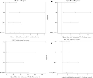
Methods This was a case–control study of women admitted in labour to one of eight hospitals of Northwell Health from January 2015 to June 2021. Objectives Examine the association between arrhythmias and adverse maternal outcomes in women with structurally normal hearts. Arrhythmia was previously diagnosed in 58.0% AF and 8.1%
This is ischemic ST depression, and could be due to increasing tachycardia, with a heart rate over 130, but that is unlikely given that the patient is now complaining of crushing chest pain and that there was tachycardia all along. There is widespread ST depression. Figure-1: Comparison of the first 2 ECGs in today's case.
Methods Patients with symptomatic PAF who underwent RFCA between October 2021 and May 2023 were retrospectively analysed. Regular clinical follow-ups were conducted to detect AF recurrence, defined as any episode of atrial fibrillation, atrial flutter or atrial tachycardia lasting >30 s.
There is a run of polymorphic ventricular tachycardia — which given the QT prolongation, qualifies as Torsades de Points ( TdP ). This patient was having recurrent episodes of polymorphic ventricular tachycardia with an underlying long QT interval ( = Torsades des Pointes ). ECG #2 Interpretation of ECG #2: Underlying sinus rhythm.
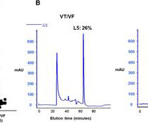
Background Early ventricular tachycardia/fibrillation (VT/VF) in patients with ST-elevation myocardial infarction (STEMI) has higher morbidity and mortality. Methods We analyzed data from 2,964 consecutive STEMI patients between January 1, 2008 and December 31, 2021.
This progressed to electrical storm , with incessant PolyMorphic Ventricular Tachycardia ( PMVT ) and recurrent episodes of Ventricular Fibrillation ( VFib ). The patient improved, and on Day-11 of the hospital stay — he was off inotropes and on a small dose of a ß-blocker. He required multiple defibrillations within a period of a few hours.
Patients with SP or AVB, 21 years of age or younger, who underwent CNA between 2015 and 2021 were included. with one documented SP after termination of atrial tachycardia at the 3-month follow-up. Objective Describe the methodology and role of CNA for treatment of pediatric patients with functional AVB or SP. The median age was 18.9
This is sinus tachycardia (rhythm) with complete heart block (AV node function) with ventricular escape rate just below 30. Never forget that sinus tachycardia is the scariest arrhythmia. The November 27, 2021 post ( LA-RA reversal ). The February 11, 2020 post ( LA-RA reversal ). The March 18, 2020 post ( LA-RA reversal ).
Be careful to differentiate bifid T waves from other pathologic conditions such as hypokalemia (which can cause a U wave with prolonged QU interval) or non-conducted P waves hidden within the T wave. == MY Comment by K EN G RAUER, MD ( 7/10/2021 ): == Our appreciation to Dr. Runyon for this case.
ACUTE MI (I allowed Acute MI to be in the report because I knew there would be an elevated troponin from ischemia, which is the definition of acute MI -- but in this case it would most likely be a Type 2 MI from tachycardia) There is also LA-RA lead reversal. The November 27, 2021 post ( LA-RA reversal ).
2] But there is also Sinus Tachycardia! This is critical for the EMS provider, or ED clinician, as identification of Grade I ischemia (aka, HATW’s) addresses the culprit lesion at the earliest opportunity with excellent downstream prognosis for the patient. [2] Even the preserved QRS duration is at risk of imminent death. & Schocken, D.
After initiating treatment for hyperkalemia, repeat ECG showed resolution of Brugada pattern: The ECG shows sinus tachycardia. A Very Wide Complex Tachycardia. The November 27, 2021 post ( LA-RA reversal ). He also received insulin with D50, sodium bicarbonate, and kayexalate for hyperkalemia. What is the Rhythm?
(Ken Grauer points out that this 5th beat appears to be due to an early atrial beat and that these early beats continue for a few beats, suggesting a short run of atrial tachycardia.) and the 2021 YouTube Review by ICU Advantage on "Temporary Pacemakers: Modes and Basic Settings".
pre-existing, stable atherosclerosis) amidst any state of global duress – to include hypertension, hypoxia, tachycardia, hypotension, sepsis, and GI bleed, for example. There may even be significant overlap between these factors. The patient was found to be hypertensive and treated accordingly. Journal of Electrocardiology, 61 ; 41-46. [3]
Abstract Aims This prospective, cross-sectional study aimed to identify sex-based differences in diagnostic and symptom experiences in postural orthostatic tachycardia syndrome (POTS). Health-related quality of life was assessed using the EuroQol 5 Dimension tool.
2021;Available from: [link] Other references: Lindow T, Mokhtari A, Nyström A, Koul S, Smith SW, Ekelund U. Harry Mond — that outlines a user-friendly approach to — “Where Am I Pacing From?” — as can be determined based on the appearance of the 12-lead ECG ( See CardioScan — May 5, 2021 ). Ann Emerg Med [Internet].
All costs are reported in 2021 US dollars. The mean cost per hospital discharge was the highest for peripheral vascular disease ($33 700 [95% CI, $33 300–$34 000]) and ventricular tachycardia/ventricular fibrillation ($32 500 [95% CI, $32 100–$32 900]).
There was never ventricular fibrillation (VF) or ventricular tachycardia (VT), no shockable rhythm. Here is a similar case: Collapse, Ventricular Tachycardia, Cardioverted, Comatose on Arrival. Agitation, Confusion, and Unusual Wide Complex Tachycardia. There is sinus tachycardia at ~115/minute.
During observation in the ED the patient had multiple self-terminating runs of Non-Sustained monomorphic Ventricular Tachycardia (NSVT). This patient very likely has some form of idiopathic ventricular tachycardia. Of the ventricular outflow tract tachycardias (RVOT-VT) makes up 80-90%.
We organize all of the trending information in your field so you don't have to. Join thousands of users and stay up to date on the latest articles your peers are reading.
You know about us, now we want to get to know you!
Let's personalize your content
Let's get even more personalized
We recognize your account from another site in our network, please click 'Send Email' below to continue with verifying your account and setting a password.


Let's personalize your content