This site uses cookies to improve your experience. To help us insure we adhere to various privacy regulations, please select your country/region of residence. If you do not select a country, we will assume you are from the United States. Select your Cookie Settings or view our Privacy Policy and Terms of Use.
Cookie Settings
Cookies and similar technologies are used on this website for proper function of the website, for tracking performance analytics and for marketing purposes. We and some of our third-party providers may use cookie data for various purposes. Please review the cookie settings below and choose your preference.
Used for the proper function of the website
Used for monitoring website traffic and interactions
Cookie Settings
Cookies and similar technologies are used on this website for proper function of the website, for tracking performance analytics and for marketing purposes. We and some of our third-party providers may use cookie data for various purposes. Please review the cookie settings below and choose your preference.
Strictly Necessary: Used for the proper function of the website
Performance/Analytics: Used for monitoring website traffic and interactions
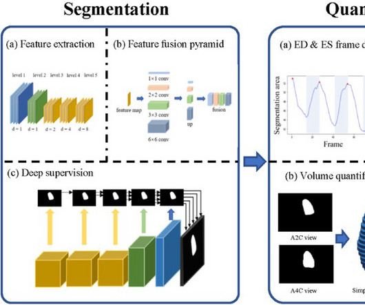
By applying AI to echocardiograms, we can help clinicians more easily detect the signs of heart valve disease so that patients get the care they need as soon as possible. Investigators trained a deep-learning program to flag patterns of tricuspid regurgitation in 47,312 echocardiograms done at Cedars-Sinai between 2011 and 2021.
Echocardiogram was performed for patients with ECMO, including at pre-ECMO, during cannulation, during ECMO support, during the ECMO wean, and a follow up within 3 months after weaning.
The algorithm uses deep learning to analyse routine ultrasound scans of the heart ( echocardiograms ) to detect disease that often goes undetected during standard assessments.
See this post: What do you think the echocardiogram shows in this case? Previously placed stents in the LAD (multiple) and mid circumflex and patent Formal echocardiogram: Normal left ventricular size and wall thickness. Shortly thereafter , the troponin came back at 3,129 ng/L (very high).
males), referred for a stress echocardiogram (SE), who underwent ESE between July 2020 (immediate post lockdown) and January 2021 according to national safety guidelines, in addition to patients wearing masks during ESE. Methods and results Baseline data were collected prospectively on 740 consecutive patients (mean age 61.4
from March 2020 to October 2021. Patients were included if they had: 1) acute COVID-19 infection confirmed by RT-PCR and 2) a transthoracic echocardiogram (TTE) performed during their hospitalization. Adult patients were identified by hospitalizations using ICD-10 code U07.1 male) were included.
The patient presented again in 2021 with right facial paresthesia and arm weakness. Repeat echocardiograms did not show recurrence of a myxoma. The patient’s past medical history was pertinent for bilateral occlusion of the femoral and popliteal arteries, with workup revealing a left atrial myxoma that was surgically resected in 2005.
Our previous research revealed that PFO shunt enables the accumulation of homocysteine in circulation (Deng, Neurology 2021). Residual shunt post PFO closure was assessed using transthoracic echocardiogram (TTE) with saline contrast. However, the mechanism underlying this association remains poorly understood.
And following the valve implantation procedure, doctors assess the valve’s integrity and blood flow control through an echocardiogram. Before implantation, doctors can adjust the valve diameter to match the patient’s heart anatomy.
We believe they are likely a normal variant in this context, and the study above failed to identify any clinically significant finding after exam and echocardiogram in 110 children with bifid T waves. sec and voltage greater than or equal to 0.05
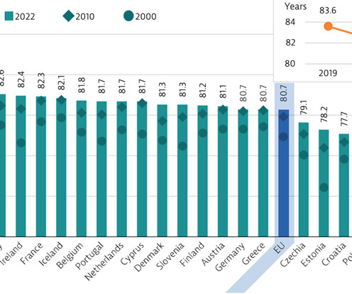
For instance, the average waiting time for an echocardiogram at Turin’s Molinette Hospital was 31 days in 2016 and an even longer 53 days for a Holter ECG. The Covid-19 emergency in 2021 accelerated the regulatory shift by highlighting the need not to overload hospital structures as well as the opportunities offered by digital healthcare.
Formal Echocardiogram: Normal left ventricular size and wall thickness. For example — Baldi et al note a more than doubling in the number of false-positive ECGs for STEMI i f judgments were based on post-resuscitation 12-lead tracings obtained less than 7 minutes after ROSC ( Resuscitation 162:445-446, 2021 ).
His echocardiogram showed normal wall motion. Figure-2: Classification of Underlying Diagnoses in Patients with MINOCA ( Adapted from Table-1 in Sykes et al: Interventional Cardiology Review: 16:e10, 2021 ). The patient did well afterward without any recurrence of symptoms. There are no further EKGs or troponin measurements.
Next day echocardiogram showed inferolateral hypokinesia with an EF of %45-50. On echocardiogram you will not see a "posterior" hypokinesia (will see "inferolateral") and, as in this case, LCx may not give the blood supply of basal inferior segment (formerly called "posterior"). 2021 Dec 7;10(23):e022866. Epub 2021 Nov 15.
Formal Echocardiogram: The estimated left ventricular ejection fraction is 58 %. Epub 2021 Nov 17. Because it reperfused on its own and because we intervened before it could re-occlude. Here is the post intervention ECG: Reperfusion T-waves identical to Wellens' syndrome, because it is the same pathophysiology. Am J Emerg Med.
Here is the cath report: Echocardiogram: There is severe hypokinesis of entire LV apex and apical segment of all the walls. Figure-2: Classification of Underlying Diagnoses in Patients with MINOCA ( Adapted from Table-1 in Sykes et al: Interventional Cardiology Review: 16:e10, 2021 ). ng/mL by 4th generation and older assays.)
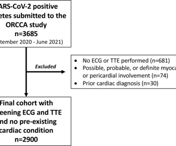
Methods The Outcomes Registry for Cardiac Conditions in Athletes was a nationwide prospective multicentre observational cohort study that captured testing and outcomes data from 45 institutions (September 2020–June 2021). Athletes with an ECG and transthoracic echocardiogram (TTE) and no pre-existing conditions were included.
He visited an outpatient clinic for it and an echocardiogram and exercise stress test was normal. In the meantime, cardiology consultant sees the patient and performs a bedside echocardiogram which revealed no major wall motion abnormalities. 2021 Sep;49(6):488-500. He has 40 packs-year of smoking history. doi: 10.5543/tkda.2021.21026.
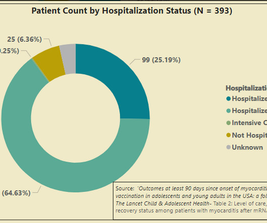
In August 2021, the CDC began a study to follow-up cases of myocarditis in the age group at highest risk for myocarditis after mRNA COVID-19 vaccination. I have to make the obligatory post-script here that I oversaw the administration of hundreds of mrna vaccines starting in March of 2021 in my cardiology clinic.
He received his first dose of a Pfizer mRNA COVID19 vaccine in June 2021. His cardiac testing completed to date consist of an electrocardiogram and an echocardiogram performed Feb 16th, 2023 that were both normal. What follows is data produced since the vaccine rollout that is relevant to Mr. Smith’s case.
Christopher Orozcos journey began on August 6, 2021, when he entered the world full of hope and love, though his story would soon take an unexpected turn. An echocardiogram revealed the heart condition that would define much of his early life: severe congenital heart defects.
It is reasonable to perform an echocardiogram to evaluate LV function. Catheter ablation or flecainide should be considered in symptomatic patients with idiopathic VT/PVCs from an origin other than the RVOT or the left fascicles. [ 1 ] Considerations Regarding Use of Flecainide: A 12-lead ECG is mandatory before starting therapy.
An echocardiogram at 13:40 showed: Severely reduced global systolic function with an estimated EF of 10-20% Mildly increased LV size Akinesis of the entire septum and apex Hypokinesis of the anterior, anterolateral, and mid posterior segments A final troponin T was drawn at 17:23- 3,475 ng/L.
We organize all of the trending information in your field so you don't have to. Join thousands of users and stay up to date on the latest articles your peers are reading.
You know about us, now we want to get to know you!
Let's personalize your content
Let's get even more personalized
We recognize your account from another site in our network, please click 'Send Email' below to continue with verifying your account and setting a password.


Let's personalize your content