This site uses cookies to improve your experience. To help us insure we adhere to various privacy regulations, please select your country/region of residence. If you do not select a country, we will assume you are from the United States. Select your Cookie Settings or view our Privacy Policy and Terms of Use.
Cookie Settings
Cookies and similar technologies are used on this website for proper function of the website, for tracking performance analytics and for marketing purposes. We and some of our third-party providers may use cookie data for various purposes. Please review the cookie settings below and choose your preference.
Used for the proper function of the website
Used for monitoring website traffic and interactions
Cookie Settings
Cookies and similar technologies are used on this website for proper function of the website, for tracking performance analytics and for marketing purposes. We and some of our third-party providers may use cookie data for various purposes. Please review the cookie settings below and choose your preference.
Strictly Necessary: Used for the proper function of the website
Performance/Analytics: Used for monitoring website traffic and interactions
Cardiovascular ultrasound has played a key role in the evolution of early diagnosis of structural heart disease, led by a technology pioneered by Philips: the ‘transesophageal echocardiography’ (TEE) ultrasound transducer. Published 2020 Sep 4. [4] Diagnostics (Basel). 2020;10(9):671. J Am Coll Cardiol. 2020;75(25):3164-3173.
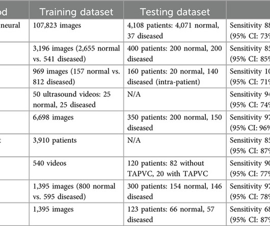
Artificial intelligence (AI)-powered ultrasound provides a potential solution to improve the diagnostic accuracy of fetal CHD screening.MethodsA literature search was conducted across seven databases for systematic review. Articles were retrieved based on PRISMA Flow 2020 and inclusion and exclusion criteria.
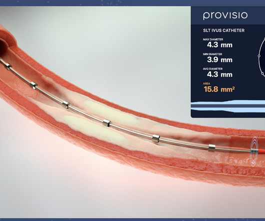
The SLT IVUS Support Crossing Catheter is an over-the-wire intravascular ultrasound catheter with an ultrasound transducer array at the distal end that also functions as a support crossing catheter. Intravascular Ultrasound Imaging Versus Digital Subtraction Angiography in Patients with Peripheral Vascular Disease.
BACKGROUND:Prior clinical trials have demonstrated the efficacy of ultrasound-facilitated catheter-directed thrombolysis (USCDT) for the treatment of acute intermediate-risk pulmonary embolism (PE) using reduced thrombolytic doses and shorter infusion durations. Circulation: Cardiovascular Interventions, Ahead of Print.
This study aims to investigate the relationship between sdLDLC level and PP in patients with stable coronary artery disease.MethodsWe conducted a retrospective analysis of 146 lesions in 86 patients by repeat intravascular ultrasound examinations from January 2020 to May 2023.
Ultrasound techniques currently used in echocardiography uses frame rates from 30-150 frames/s. While conventional ultrasound uses focused beam transmission, ultrafast ultrasound uses unfocused plane-wave ultrasound which can result in very high temporal resolution with frame rates up to 100 times faster [2]. 2019.09.019.
We used carotid ultrasounds to detect plaque at baseline and follow‐up in 2006 to 2009 (median follow‐up=5.5 We identified incident CVD events through 2020 with a median follow‐up of 18.5 had incident plaque (109/1104 plaque‐free participants with baseline and follow‐up ultrasounds), 11.0% Approximately 2.8%
The incidence of no-reflow was higher in patients with attenuated plaque ≥5 mm in length as evaluated by intravascular ultrasound (IVUS).Objective:The The incidence of no-reflow was higher in patients with attenuated plaque ≥5 mm in length as evaluated by intravascular ultrasound (IVUS).Objective:The
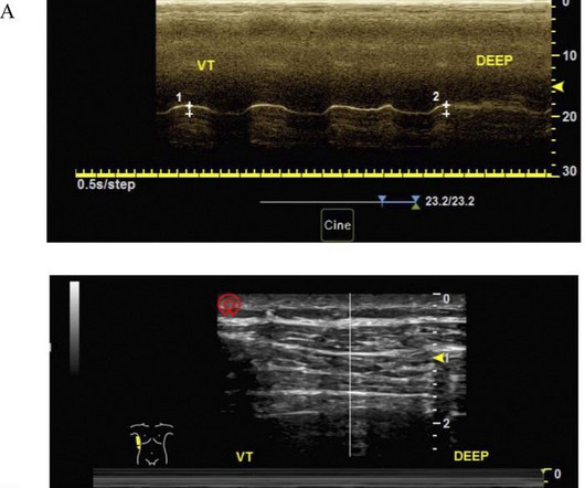
Diaphragmatic dysfunction was defined as a diaphragmatic excursion of less than 9 mm in women and less than 10 mm in men at rest, with an average thickening fraction of less than 20%.
A retrospective single centre cohort analysis of attempted fetal aortic valvuloplasty from a pioneering centre, of cases from 2000 to 2020 has been published [1]. Currently all procedures are done percutaneously under ultrasound guidance following maternal spinal or epidural anaesthesia. Reference Ryan Callahan, Kevin G. Esch, Lynn A.
Before the procedure, patients should have an electrocardiogram (ECG) and echocardiogram (ultrasound of the heart) to check the heart’s rhythm and function. 2020 ESC Guidelines for the diagnosis and management of atrial fibrillation developed in collaboration with the European Association of Cardio-Thoracic Surgery (EACTS).
IVI was defined as intravascular ultrasound or optical coherence tomography. A contemporary subset of PCIs from 2020 to 2022 was used to examine clinical characteristics associated with IVI use and test the reliability of IVI as a pass/fail performance measure.
A formal ultrasound later showed reasonably good LV function, and so he later received carvedilol and diltiazem, Unfortunately, those led to hypotension at 80/40 with a HR 40. Several hours later, this was the effect: NT pro-BNP elevated to 7000 Furosemide was also given. I'd add the following thoughts to Dr. Smith's discussion.
The updated workflow indicates that direct imaging guidance, such as ultrasound, may be used as an alternative to fluoroscopy. "As 2020 June;127(1):4-20. This is the most commonly used ablation catheter in the world for RF ablation and is fully integrated with the CARTO 3 System. Int J Stroke. 2021 Feb;16(2):217-221.
24: Joint American College of Cardiology/Journal of the American College of Cardiology Late-Breaking Clinical Trials (Session 402) Saturday, April 6 9:30 – 10:30 a.m.
This was a point of care ultrasound, not a bubble contrast echo. On echo, OMI has a wall motion abnormality, but SI usually does not. == MY Comment by K EN G RAUER, MD ( 5/13/2020 ): == It’s good to periodically review the differential diagnosis of the diffuse subendocardial ischemia. What do you think the echocardiogram shows?
Case continued A bedside ultrasound showed diminished LV EF and of course bradycardia. The February 11, 2020 post ( LA-RA reversal ). The March 18, 2020 post ( LA-RA reversal ). The August 28, 2020 post ( LA-LL reversal ). The November 19, 2020 post ( LA-LL reversal ). RVMI explains part of the shock.
The primary efficacy outcome was the composite of no occlusion in the treated segment assessed at serial duplex ultrasound examinations or no reintervention needed to maintain patency within 6 months. Secondary outcomes, including Villalta score, quality of life, and safety outcomes, were also assessed.
Further ultrasound showed no B-lines (no pulmonary edema). 23/WCC — 2/21/2020 ). 23/WCC — 2/21/2020 ). 23/WCC — 2/21/2020 ). 23/WCC — 2/21/2020 ). There is very little filling, and thus very poor stroke volume. The heart rate is too fast for this poor filling.
To address this issue, we propose a novel approach using Superb Microvascular Imaging (SMI), an ultrasound method that that enables detailed visualization of microvascular flow, to assess renal microcirculation.Methods:We retrospectively analyzed 78 patients who underwent renal ultrasonography using SMI from October 2020 to May 2023.
Another approach is sympathetic chain (stellate ganglion) blockade if you have the skills to do it: it requires some expertise and ultrasound guidance. As I review in My Comment in the January 16, 2020 post of Dr. Smith's ECG Blog ( and have reproduced in Figure-2 below ) — the ECG of patients with acute LMCA occlusion may be varied.
On arrival, lung ultrasound confirmed pulmonary edema (B lines). In comparison to the previous study, 11/11/2020, there has been a significant interval deterioration of left ventricular systolic function (previous EF 80%), and there is a new large apical wall motion abnormality. This is proximal LAD Occlusion until proven otherwise.
EKG from triage: Here is his previous ECG: Normal ST Elevation Resident's interpretation: Reperfusion pattern/Wellens' with biphasic T waves in V2 and V3, and in comparison to an EKG in 2020 this is new. Bedside ultrasound with no apparent wall motion abnormalities, no pericardial effusion, no right heart strain.
The ways to tell for certain include intravascular ultrasound (to look for extra-luminal plaque with rupture) or "optical coherence tomography," something I am entirely unfamiliar with. The authors recommend using optical coherence tomography or intravascular ultrasound imaging in patients with evidence of nonobstructive CAD by angiogram.
A bedside cardiac ultrasound was normal, with no effusion. Clin Chem [Internet] 2020;Available from: [link] Smith mini-review: Troponin in Emergency Department COVID patients Cardiac Troponin (cTn) is a nonspecific marker of myocardial injury. JAMA Cardiol [Internet] 2020;Available from: [link] 4. Guo T, Fan Y, Chen M, et al.
After rethinking the case, he remained concerned about ACS and subsequently performed a point-of-care ultrasound in order to evaluate for regional wall motion abnormality. In equivocal cases, point-of-care ultrasound may be the difference between taking the patient to the lab or not. 1] Wereski, R., Chapman, A. Gray, A., &
The problem is difficult to study because angiographic visualization of arteries is not perfect, and not all angiograms employ intravascular ultrasound (IVUS) to assess for unseen plaque or for plaque whose rupture and ulceration cannot be seen on angiogram. Thus, intracoronary imaging modalities are crucial in this setting.
Two thirds of MINOCA cases are due to atherosclerotic causes One way to prove the diagnosis in this case would have been with intravascular imaging such as optical coherence tomography (OCT) or intravascular ultrasound (IVUS). Fortunately, that is exactly what happened. This is not the case.
Billion in 2020. Magnetic Resonance-Guided Ultrasound It is an MRI-based therapeutic technique that makes use of ultrasonic pulses to remove the target tissue. It has greatly enhanced the methods of diagnostics and has made the treatment of several medical conditions more efficient and effective.
The sheep were anesthesitized and handled under approval of the Animal Research Committee at UCLA (protocol ARC 2020‐19). Femoral artery and external jugular vein access were obtained using the Seldinger technique under ultrasound guidance and did not require a surgical cut down.
In 2020, the state medical board investigated Dr. Mustapha and referred him to the Michigan attorney general. A lower extremity arterial ultrasound revealed elevated velocities in the right proximal superficial femoral artery. A second opinion from another physician at another facility resulted in a normal ultrasound.
This case was provided by Spencer Schwartz, an outstanding paramedic at Hennepin EMS who is on Hennepin EMS's specialized "P3" team, a team that receives extra training in advanced procedures such as RSI, thoracostomy, vasopressors, and prehospital ultrasound. Takotsubo is a sudden event, not one with crescendo angina.
I would do bedside ultrasound to look at the RV, look for B lines as a cause of hypoxia (which would support OMI, and argue against PE), and if any doubt persists, a rapid CT pulmonary angiogram. As for the ECG, it could represent OMI, but RBBB is also a clue that it may be PE.
Dr. Nossen performed a bedside ultrasound which was interpreted as normal. As emphasized in My Comment at the bottom of the page of the June 3, 2020 post in Dr. Smith's ECG Blog — Almost everyone gets fooled the 1st time they see this phenomenon. The KEY to interpreting today's case — is to be aware of the Emery Phenomenon.
No pericardial effusion on ultrasound." Smith's ECG Blog how difficult it may sometimes be to distinguish between acute myocarditis vs acute OMI on the basis of ECG findings and the clinical history ( See My Comments in the July 21, 2019 — December 10, 2019 — and January 10, 2020 posts). What do you think?
A bedside ultrasound should be done to assess volume and other etiologies of tachycardia, but if no cause of type 2 MI is found, the cath lab should be activated NOW. The November 10, 2020 post — for PTA. The October 17, 2020 post — for a 70-year old woman with " Artifactual VT ". The March 17, 2023 post — for PTA.
A bedside cardiac ultrasound was performed with a parasternal long axis view demonstrated below: There is a large pericardial effusion with collapse of the right ventricle during systole. Figure reproduced from My Comment at the bottom of the page in the September 7, 2020 post in Dr. Smith's ECG Blog ). She has already had syncope.
In other words, I believe this ultrasound result refers to the lateral wall that is recorded by I, aVL, V5, V6; terminology has changed such that anything that is either posterior or lateral is now called "lateral."
Here is the parasternal short axis, performed by a real expert in emergency department point of care cardiac ultrasound: There does not appear to be an anterior wall motion abnormality. Beware a negative Bedside ultrasound. Exam revealed no friction rub What do you want to do? A bedside echo was done immediately. Pericarditis?
The vaccine efficacy data for the original data was from thousands of patients and I certainly felt given the devastation wreaked on many of my patients in 2020 that the vaccines were the best chance of avoiding morbidity and mortality.
Smith comment: Point of Care ultrasound is not adequate to rule out wall motion abnormality; moreover, diffuse subendocardial ischemia often has no wall motion abnormality because the epicardium is still contracting. Association between opioid analgesia and delays to cardiac catheterization of patients with occlusion Myocardial Infarctions.
Regional wall motion abnormality-inferolateral (this is the formal ultrasound location of a posterior wall motion abnormality). Formal contrast echo: The estimated left ventricular ejection fraction is 44%. Regional wall motion abnormality-anterolateral. Is the ability to diagnose posterior reperfusion a useful skill? 2022.08.750 Section 5.2.2,
So I immediately left the room to get an ultrasound machine. While calling for some help and arranging to have her transported to our critical care zone, I got this quick ultrasound which confirmed my suspicion: This quick view was all I was able to obtain in the circumstances.
This is a case written by Dan Lee (One of our fantastic Hennepin Residents, class of 2020 ) edits by Smith A 60 something-year-old man with a history of ESRD, LVH and prior CABG presented after an episode of hypotension during his hemodialysis, run followed by a syncopal episode which caused his run to be terminated early.
We organize all of the trending information in your field so you don't have to. Join thousands of users and stay up to date on the latest articles your peers are reading.
You know about us, now we want to get to know you!
Let's personalize your content
Let's get even more personalized
We recognize your account from another site in our network, please click 'Send Email' below to continue with verifying your account and setting a password.


Let's personalize your content