This site uses cookies to improve your experience. To help us insure we adhere to various privacy regulations, please select your country/region of residence. If you do not select a country, we will assume you are from the United States. Select your Cookie Settings or view our Privacy Policy and Terms of Use.
Cookie Settings
Cookies and similar technologies are used on this website for proper function of the website, for tracking performance analytics and for marketing purposes. We and some of our third-party providers may use cookie data for various purposes. Please review the cookie settings below and choose your preference.
Used for the proper function of the website
Used for monitoring website traffic and interactions
Cookie Settings
Cookies and similar technologies are used on this website for proper function of the website, for tracking performance analytics and for marketing purposes. We and some of our third-party providers may use cookie data for various purposes. Please review the cookie settings below and choose your preference.
Strictly Necessary: Used for the proper function of the website
Performance/Analytics: Used for monitoring website traffic and interactions
Background:Cerebral venous thrombosis (CVT) is associated with significant risk of death and disability. Adult patients in 2018 (n=6,051,974) were compared to those in 2020 (n=5,470,813), the first year of the COVID-19 pandemic. Hospital mortality rate among CVT patients admitted in 2020 was 5%.
Background:Coronavirus disease 2019 (COVID-19) increases the risk of cerebral venous sinus thrombosis (CVST), and previous reports derived from small case series reported a high mortality in these patients, up to 40%. Median age did not differ between the groups (43 vs 44years,P=0.6)
These included three cases of intraoperative thrombosis, three instances of pericardial effusion or tamponade, one case of device-related thrombosis, one peri-device leak, one systemic embolism, one bleeding episode, and one additional device-related complication.
The risk of stent thrombosis is particularly increased during the first 6 months after intervention. Key exclusion criteria included <18 or >75 years of age, contraindications to anticoagulant use, or acute venous thrombosis <3 months. The study was registered atClinicalTrials.gov(NCT04128956).RESULTS:From
Introduction:Seizures are a common initial manifestation of cerebral venous thrombosis (CVT). The primary outcome was delayed seizure(s), occurring after 7 days from CVT diagnosis.
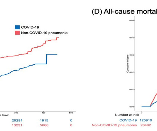
Researchers analyze primary and secondary cardiovascular outcomes in 132,784 inpatients with COVID-19 (October 8, 2020 to September 30, 2021) and 31,173 inpatients with non-COVID-19 pneumonia (January 1, 2019 to December 31, 2019) in Korea. The results indicate a lower risk of cardiovascular disease in COVID-19 patients.
Principal safety outcomes were independently adjudicated International Society on Thrombosis and Haemostasis major bleeding at 72 hours post-treatment and mortality within 12 months of treatment.
MINOCA may be due to: coronary spasm, coronary microvascular dysfunction, plaque disruption, spontaneous coronary thrombosis/emboli , and coronary dissection; myocardial disorders, including myocarditis, takotsubo cardiomyopathy, and other cardiomyopathies. This is in spite of the known proclivity of tighter stenoses to thrombose.
We sought to explore the relationship between ART and stroke risk using population-level data.Methods:We conducted a retrospective cohort study using data from the National Inpatient Sample (NIS) registry from 2015-2020, including all delivery hospitalizations for patients aged 15-55 years. The study exposure was use of ART.
There is a literature on this subject ( Sood et al — Cureus 15(4):e37102, 2023 — Gulati et al — Mayo Clin Proceed 95(1):136-156, 2020 — GGF van der Schoot et al: Neth Heart J 28(6):301-308, 2020 — and — Egred at al — Postgrad Med 81(962): 741-745, 2005 — to name just a few reports ). That said — acute MI does occur in younger patients.
In severe OHSS, increases in capillary permeability can result in hemoconcentration and hypercoagulability leading to thrombotic events, including stroke and cerebral venous thrombosis. HCUP contains both emergency department and inpatient encounters whereas NIS contains a nationally representative sample of inpatient encounters.
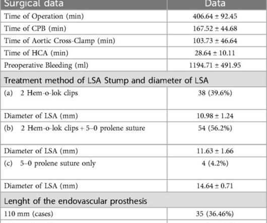
ObjectiveThis study aims to summarize the clinical experience of using Hem-o-lok clips for the closure of the left subclavian artery (LSA) stump in patients with acute Stanford type A aortic dissection.MethodsClinical data were collected from 96 patients with acute type A aortic dissection admitted to our hospital from January 2020 to December 2022.
The present study aims to characterize racial disparities in long-term outcomes, perioperative outcomes, and health care use after endovascular aortic aneurysm repair.METHODS:We conducted a retrospective cohort study from 2011 to 2019 with outcome assessment through 2020.
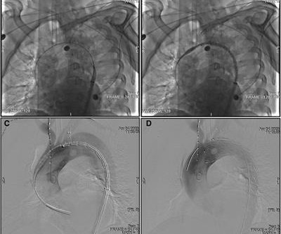
This study investigates clinical outcomes, aortic remodeling, and abdominal aortic perfusion patterns after TEVAR with the novel Castor device.MethodsFrom November 2020 to June 2023, 29 patients with TBAD involving the LSA were treated with the Castor single-branched stent graft. Of the 102 abdominal aortic branches, 94.1%
The 2020 European Society of Cardiology (ESC) Guidelines recommend the use of prasugrel over ticagrelor in patients with non-ST-elevation ACS (NSTE-ACS) intended to receive invasive management (class IIa recommendation), however there are contradictory views regarding this recommendation.
We who know ischemic ECGs know that really when T-wave inversion is specific for coronary thrombosis that it indicates reperfusion of the artery, not active occlusion. For examples of this phenomenon — See My Comment in the February 14, 2018 — July 21, 2020 — and December 22, 2022 posts in Dr. Smith's ECG Blog ).
Based on the data of discharge patients from 2015 to 2020 (Model Training Set), logistic regression model with restricted cubic splines were applied to construct the "prognostic scale of AIS acute stage based on treatment stratification, PAIST Scale". Patients treated with arterial thrombectomy were excluded. mmol/L = 1).
Methods Patients from a large US hospital system undergoing combined LAAO and left-atrial CA from 8/2020 to 2/2024 were retrospectively analyzed and compared to a control group undergoing LAAO alone. vs. 30.4%, p =0.07) and device related thrombosis (4.5% vs. 2.1%, p =0.72) and minor (27.8%
With the information, the APN would recommend change in management accordingly after communicating with the neurology team from the original transferring acute hospital.Results:There were total of 51 issues identified from Aug 2020 to Aug 2021 from 125 patients were reviewed by the Stroke APNs.
The World Health Organisation (WHO) formally proclaimed COVID-19, the illness spread by a zoonotic SARS-CoV-2, as a pandemic in March 2020, after it had started to spread in late 2019. Even though the pandemic is on the wane, new studies and evidence about it continue to emerge.
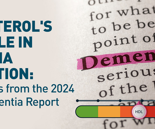
Updated with the latest research since the 2020 edition, this comprehensive report underscores the potential to prevent or significantly delay dementia by targeting modifiable risk factors. Key Takeaways: Up to 50% of all dementia cases could potentially be prevented or substantially delayed.
We aim to assess how AI-ECG prediction model outputs, specifically the AF probability and delta age, are associated with adverse vascular outcomes in patients with migraine.Adult patients diagnosed with MwA and MwoA from 2000-2020 with at least one digital, standard 12-lead ECG were identified.
In the early years of percutaneous coronary intervention (PCI), studies indicated a heightened risk of major adverse cardiac events (MACE) in patients with reduced left ventricular ejection fraction (LVEF), involving outcomes such as death, Q-wave myocardial infarction (MI), stent thrombosis, and repeat revascularization.
MINOCA may be due to: coronary spasm, coronary microvascular dysfunction, plaque disruption, spontaneous coronary thrombosis/emboli , and coronary dissection. link] We know that most type 1 acute MI due to plaque rupture and thrombosis occurs in lesions that are less than 50% (see Libby reference).
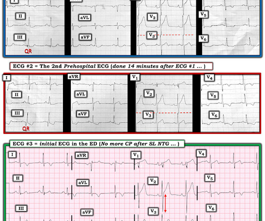
A second 12 Lead ECG was recorded: This is a testament to the dynamic nature of coronary thrombosis and thrombolysis. The patient verbalized spontaneous improvement just before 324mg ASA administration. Here the ST segments are not so deep, nor are the T waves so wide and bulky, because of improved coronary flow at the level of the occlusion.
Microvascular dysfunction — cardiac vasculitis — intravascular thrombosis. These cases provide insight to assessment for MAT: The January 5, 2020 post in Dr. Smith’s ECG Blog — for an example of MAT. Acute pulmonary embolus. ARDS ( A dult R espiratory D istress S yndrome ). SIRS ( S ystemic I nflammatory R esponse S yndrome ).
The commonest causes of MINOCA include: atherosclerotic causes such as plaque rupture or erosion with spontaneous thrombolysis, and non-atherosclerotic causes such as coronary vasospasm (sometimes called variant angina or Prinzmetal's angina), coronary embolism or thrombosis, possibly microvascular dysfunction. This is not the case.
Nevertheless, I don't think a thrombosis related type I MI was ruled out here simply because the patient refused further evaluation. IF you missed the KEY Findings on the pre-hospital ECG of todays case Please take another look at My Comment at the bottom of the page of that February 6, 2020 post.
Coronary thrombosis or embolism can result in MINOCA, either with or without a hypercoagulable state. Shark Fin morphology has been discussed a number of times on Dr. Smith’s ECG Blog ( For review — See the June 11, 2018 post and the January 24, 2020 post , to name just 2 instances ). 24, 2020 post ( link to that post given above ).
12,16 In 2017, CANTOS (Canakinumab Anti-inflammatory Thrombosis Outcomes Study) provided proof-of-principle that inflammation inhibition in the absence of lipid lowering can significantly reduce cardiovascular event rates and helped to define the interleukin-1 (IL-1) to IL-6 to CRP pathway as a central target in CV disease.16 Circulation.
There is a literature on this subject ( GGF van der Schoot et al: Neth Heart J 28(6):301-308, 2020 — and — Egred et al — Postgrad Med 81(962): 741-745, 2005 — to name just 2 reports ). Meyers at the end of his discussion that refer to multiple cases published on Dr. Smith’s ECG Blog of young adults with acute MI ). Was this coincidence? —
The reappearance of de Winter's pattern caused by acute stent thrombosis: A case report. Academic Emergency Medicine 27(S1): S220; May 2020. Prominent T wave and J-point depression in the precordial leads associated with ST-segment elevation in lead aVR. Am J Emerg Med. 2014;32:e5–e8. Hayakawa A, Tsukahara K, Miyagawa S, et al.
This is a case written by Dan Lee (One of our fantastic Hennepin Residents, class of 2020 ) edits by Smith A 60 something-year-old man with a history of ESRD, LVH and prior CABG presented after an episode of hypotension during his hemodialysis, run followed by a syncopal episode which caused his run to be terminated early.
IntroductionCerebral venous thrombosis (CVT) is an uncommon form of stroke with relatively low mortality but higher incidence in younger adults.1–3 IntroductionCerebral venous thrombosis (CVT) is an uncommon form of stroke with relatively low mortality but higher incidence in younger adults.1–3 in univariable regression. vs APC 6.8%).
IntroductionCerebral Venous Thrombosis (CVT) is a rare cerebrovascular condition causing death or functional dependency in 10‐15%. Stroke: Vascular and Interventional Neurology, Volume 3, Issue S2 , November 1, 2023. CVT and other diagnoses, and EVT and other procedures, were identified using standard ICD 10 codes.
Introduction:Active cancer is a known risk factor for cerebral venous thrombosis (CVT), but it is unknown whether CVT is also associated with occult cancer. Patients admitted to any Dutch hospital between 1997 and 2020 with a first episode of CVT were identified using ICD-9 and ICD-10 codes.
Introduction:COVID-19 infection has thus emerged to be a new risk factor for Cerebral Venous Thrombosis (CVT). Methods:Adult patients with CVT diagnosis from 2020-2022 in TriNetX COVID research network were included in the study. CVT with COVID patients were more likely to develop deep vein thrombosis at one month (19.8%
The composition of thrombi may reflect the mechanism of thrombosis, aiding the determination of the treatment strategy. There was a positive correlation between thrombin and platelets in the cancer group (r=0.666;P=0.001) but not in the control group (r=−0.167;P=0.627).CONCLUSIONS:Cerebral
Background and Purpose:Cancer increases the risk for acute ischemic stroke (AIS) and deep venous thrombosis. Methods:We included AIS patients hospitalized at our comprehensive stroke center between January 2015 and December 2020 with available PFO status as detected on transesophageal echocardiography.
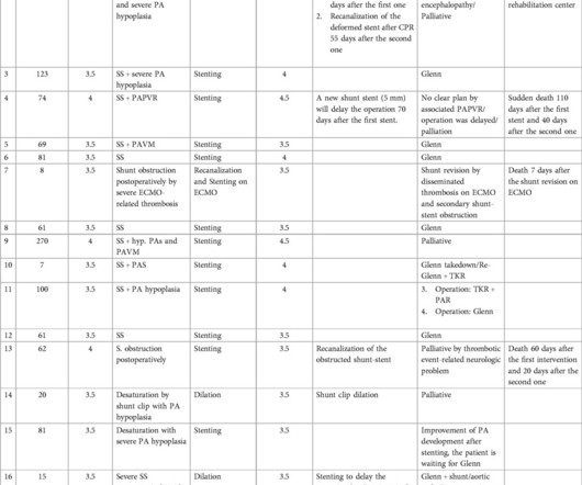
MethodsThis retrospective study was conducted between 2020 and 2024, when 24 urgent interventions were performed on 17 patients at our center. However, managing late complications such as stent thrombosis remains a significant challenge. kg), respectively. Notably, there were no intervention-related complications or deaths.
We organize all of the trending information in your field so you don't have to. Join thousands of users and stay up to date on the latest articles your peers are reading.
You know about us, now we want to get to know you!
Let's personalize your content
Let's get even more personalized
We recognize your account from another site in our network, please click 'Send Email' below to continue with verifying your account and setting a password.


Let's personalize your content