This site uses cookies to improve your experience. To help us insure we adhere to various privacy regulations, please select your country/region of residence. If you do not select a country, we will assume you are from the United States. Select your Cookie Settings or view our Privacy Policy and Terms of Use.
Cookie Settings
Cookies and similar technologies are used on this website for proper function of the website, for tracking performance analytics and for marketing purposes. We and some of our third-party providers may use cookie data for various purposes. Please review the cookie settings below and choose your preference.
Used for the proper function of the website
Used for monitoring website traffic and interactions
Cookie Settings
Cookies and similar technologies are used on this website for proper function of the website, for tracking performance analytics and for marketing purposes. We and some of our third-party providers may use cookie data for various purposes. Please review the cookie settings below and choose your preference.
Strictly Necessary: Used for the proper function of the website
Performance/Analytics: Used for monitoring website traffic and interactions
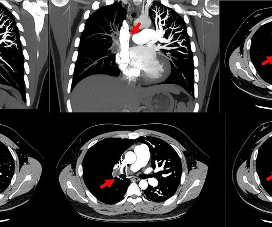
Pulmonary embolism is the most common cardiovascular disease after myocardial infarction and stroke. Konstantinides (Eur Heart J 41(4):543–603, 2020) Current guidelines categorize patients with PE as being at.
This study aims to describe a 20-year experience of pulmonary artery banding at a tertiary care center in a developing country.MethodsThis is a retrospective chart review of patients who underwent pulmonary artery banding over a 20-year period between January 2000 and July 2020 in a tertiary care center in a developing country.
BACKGROUND:Prior clinical trials have demonstrated the efficacy of ultrasound-facilitated catheter-directed thrombolysis (USCDT) for the treatment of acute intermediate-risk pulmonary embolism (PE) using reduced thrombolytic doses and shorter infusion durations.
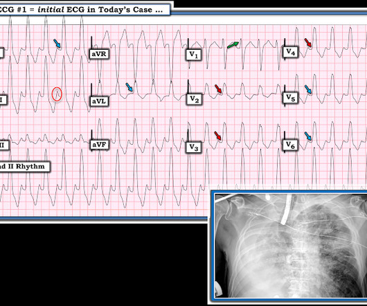
KEY Point: Although true that patients with longstanding, severe pulmonary disease may manifest a QRST complex in standard lead I with marked overall reduction in QRST amplitude ( See ECG Blog #65 — regarding Schamroth’s Sign ) — you should never normally see a completely flat line in any of the standard limb leads.
BackgroundRecurrent pulmonary vein stenosis (PVS) following surgical repair of total anomalous pulmonary venous connection is associated with poor prognosis. Since August 2020, patients have been considered for valsartan therapy early after operation.
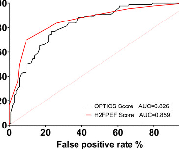
Objective Group II pulmonary hypertension (PH) can be challenging to distinguish from Group I PH without proceeding to right heart catheterisation (RHC). The diagnostic accuracy of the H2FPEF and OPTICS scores was investigated in Scotland. The diagnosis from the scores were compared with the MDT consensus diagnosis following RHC.
BackgroundIn recent years, self‐expanding technology to treat pulmonary regurgitation in the native right ventricular outflow tract became Food and Drug Administration approved in the United States and is now routinely used. Journal of the American Heart Association, Ahead of Print.
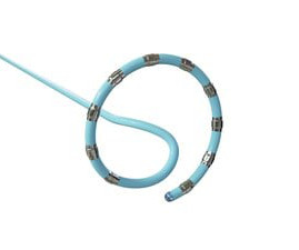
The VARIPULSE Platform is designed to enable pulmonary vein isolation with the versatility of a catheter loop, a simple generator user interface, and a mapping system that provides an intuitive, reproducible workflow with real-time visualization, contact indicator, and PF tagging mechanisms. Epub 2020 Jan 19. 2020 June;127(1):4-20.
She had acute pulmonary edema on exam. On arrival, lung ultrasound confirmed pulmonary edema (B lines). In comparison to the previous study, 11/11/2020, there has been a significant interval deterioration of left ventricular systolic function (previous EF 80%), and there is a new large apical wall motion abnormality.
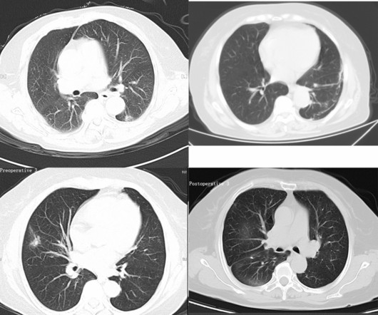
In this retrospective study, a total of 103 patients with lung cancer who received outpatient or inpatient treatment from December 2020 to May 2022 were selected and divided into the lobectomy group (n = 48) and the segmentectomy group (n = 55) according to different surgical methods.
We aimed to compare in-hospital mortality and predictors in stroke patients with secondary rheumatological conditions.Methods:Using the National Inpatient Sample (NIS), we identified patients ≥18 admitted for stroke (Jan 2019 - Dec 2020), stratified into RA, SLE, scleroderma, or vasculitides groups using ICD-10-CM codes.
BACKGROUND:The aim of this study was to examine the impact of early versus delayed catheter-based therapies (CBTs) on clinical outcomes in patients with acute intermediate-risk pulmonary embolism (PE).METHODS:This Patients were divided into early (<12 hours) and delayed CBT (12 hours) groups.
Introduction:The demographics of patients with pulmonary arterial hypertension (PAH) is shifting towards older age, increased comorbidity burden, and an increase in the risk of left ventricular (LV) diastolic dysfunction. Circulation, Volume 150, Issue Suppl_1 , Page A4146852-A4146852, November 12, 2024. 2022 were included.
In the context of today's case — these P waves are diagnostic of RAE = P Pulmonale ( See ECG Blog #75 ) and almost certain associated pulmonary hypertension. Working through a case of a regular WCT Rhythm in this 80-something woman — See My Comment in the May 5, 2020 post on Dr. Smith’s ECG Blog.
SCAPE is an acronym for sympathetic crash acute pulmonary edema, which can typcially occur in Pickering syndrome with renal artery stenosis [1]. Another term for transient acute pulmonary edema which occurs in renal artery stenosis is flash pulmonary edema. doi: 10.1136/bcr-2020-239421. Flash pulmonary edema.
The PulseSelect PFA system was engineered with differentiated safety features and provides rapid, effective pulmonary vein isolation (PVI) through consistent and predictable energy delivery and catheter maneuverability. Eur Heart J 2020. For more information: www.medtronic.com References: Verma A, et al. Wolf PA, Abbott RD, Kannel WB.
BackgroundThe modified Blalock‐Taussig‐Thomas shunt is the gold standard palliation for securing pulmonary blood flow in infants with ductal‐dependent pulmonary blood flow.
This middle-aged patient presented with SOB, weakness, and mild pulmonary edema. See this terrible case: Computer often fails to diagnose atrial fibrillation in ventricular paced rhythm, and that can be catastrophic == MY Comment , by K EN G RAUER, MD ( 1/22/2020 ): == Our THANKS to Dr. Smith for presenting this extremely interesting case.
These 2 settings are: i ) In patients with severe , often longstanding pulmonary disease ; and / or , ii ) In acutely ill patients with multi-system disease ( ie, sepsis, shock, electrolyte and/or acid-base disorders ). Hypoxic injury ( from pneumonia or other acute pulmonary complication ). Acute pulmonary embolus.
Smith interpretation: This is highly likely to be due to extreme right heart strain and is nearly diagnostic of pulmonary embolism. It is of course pulmonary embolism. No d-dimer or CT pulmonary angiogram was done when they discovered that she had normal coronary arteries. What is the clear diagnosis and reason for arrest?
All SARS-CoV-2 polymerase chain reaction (PCR) positive patients who underwent a TTE during their inpatient admission between 1 March 2020 and 31 October 2020 were analysed. A raised pulmonary artery systolic pressure (36.8%) andright ventricle (RV) dysfunction (26.4%) were the most common echocardiographic features.
ABSTRACT Introduction Pulmonary vein isolation (PVI) using a cryoballoon is well-established for the treatment of paroxysmal atrial fibrillation (PAF). Compared to other available technologies, the usage of a stable, low-pressure cryoballoon (POLARx, Boston Scientific) has demonstrated lower nadir temperatures and longer thawing times.
This patient in severe respiratory distress clearly shows sufficient abnormalities on chest X-Ray that could plausibly account for the highly unusual QRS morphology in Figure-2 ( whereas in a patient without such extensive pulmonary disease — such QRS morphology would simply not be consistent with LBBB conduction ).
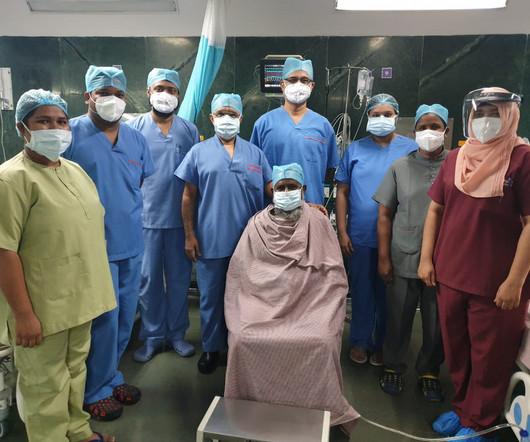
Prateek Bhatnagar, Director Cardiac Surgery & Chief Cardiology Surgeon at Care Hospital, Banjara Hills, Hyderabad, has successfully operated & discharged India’s first post Covid 19 recovered patient who on 16th July, 2020, underwent a triple bypass surgery under Dr. Bhatnagar & his team of doctors here.
Smith comment: before reading anything else, this case screamed pulmonary embolism to me. I would do bedside ultrasound to look at the RV, look for B lines as a cause of hypoxia (which would support OMI, and argue against PE), and if any doubt persists, a rapid CT pulmonary angiogram.
I've previously discussed the interesting correlation of a qR pattern in lead V1 in patients with RVH — as strongly suggesting associated pulmonary hypertension ( See ECG Blog #234 and Blog #248 ). The QRS is not wide enough for a complete RBBB — and, lateral limb leads I and aVL both lack terminal S waves.
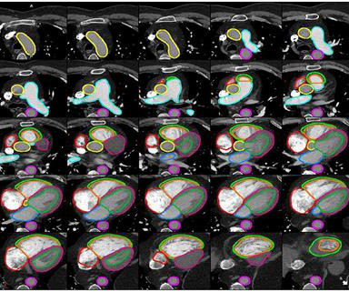
Results 18 studies published after 2020 were included. 0.92), and pulmonary artery (0.92, IQR 0.87–0.93). The DSC scores median achieved for the most commonly segmented structures were left atrium (0.88, IQR 0.83–0.91), 0.91), left ventricle (0.91, IQR 0.89–0.94), 0.94), left ventricle myocardium (0.83, IQR 0.82–0.92),
We used previously validated ICD-10-CM codes for acute ischemic stroke, intracerebral and subarachnoid hemorrhage, cerebral venous thrombosis, acute myocardial infarction, pulmonary embolism, and acute deep venous thrombosis to define our study outcome.Results:We identified a total of 747 patients with OHSS in HCUP.
Methods In a retrospective cohort study, patients who underwent isolated TVS at our institution between 2012 and 2020 were screened and followed up to 1 year. It was associated with ECHO-derived right ventricular (RV) free wall strain and RHC-derived RV systolic and diastolic as well as mean pulmonary pressures.
Leaving a fenestration in the interatrial septum during a Fontan repair is useful in relieving the central venous congestion when pulmonary blood flow is driven by venous pressure in Fontan repair. Subjects were between 2-6 years at Fontan surgery done between 2010 and 2020 with cardiac catheterization done within 1 year prior to the surgery.
The primary endpoint of the analysis was incidence of procedure – or device-related primary adverse events (PAEs) within seven to ninety days of the ablation procedure; the secondary endpoint was the rate of acute pulmonary vein (PV) isolation.1 2020 June;127(1):4-20. Int J Stroke. 2021 Feb;16(2):217-221. Benjamin EJ, Schnabel RB.
Conduction and refractoriness alternans may be seen with WPW-related as well as AV Nodal-dependent reentr y tachycardias — atrial fibrillation — acute pulmonary embolus — myocardial contusion — and severe LV dysfunction. Therefore — identification of QRS alternans during a regular SVT does not prove the existence of an accessory pathway.
The bedside echo showed a large RV (Does this mean there is a pulmonary embolism as the etiology?) When you suspect pulmonary embolism due to large RV on POCUS, always look for right axis deviation and a large R-wave in V1 because the large RV may be entirely due to chronic RVH, not acute PE. Here is his triage ECG: What do you think?
This study examines the outcomes of hospitalized AF patients with thrombocytopenia.Methods:The National Inpatient Sample (NIS) from 2016-2020 was analyzed to identify adult patients with AF and thrombocytopenia (using the proper ICD-10 codes). The primary outcome was all-cause inpatient mortality.
Methods and Results Patients with symptomatic, drug-resistant paroxysmal AF for first ablation were prospectively enrolled from September 2020 to January 2022. First-pass isolation, acute pulmonary vein (PV) reconnections, 1-year arrhythmia recurrence, and major complications were assessed.
Notes: Approved after initial rejection two years prior based on safety data from Japan, where the drug had been used since 2020 It can only be used in kidney disease patients on dialysis for at least 3 months. Clinical Trial Results: Aprocitentan reduced and maintained lower BP levels over time, both in SiSBP and SiDBP.
Methods Multicentre UK study undertaken 1 March 2020–30 June 2021 during the COVID-19 pandemic. to 1.10 (p<0.01)) and with pulmonary arterial hypertension (PAH; OR 5.99, 95% CI 1.34 Objective Ascertain the impact of COVID-19 on people with CHD and define risk factors for adverse outcomes. to 26.91 (p=0.02)).
Further ultrasound showed no B-lines (no pulmonary edema). 23/WCC — 2/21/2020 ). 23/WCC — 2/21/2020 ). 23/WCC — 2/21/2020 ). 23/WCC — 2/21/2020 ). The heart rate is too fast for this poor filling. Preload must be increased and the heart rate slowed in order to allow more LV filling.
Methods:We analyzed young adult hospitalizations (18-44 years) with known CUD from the 2020 National Inpatient Sample. vs. 22.6%), HTN (17% vs. 14.7%), chronic pulmonary disease (13.2% vs. 27.9%), visiting rural hospitals (13.9% vs. 1.3%), with Medicaid (56.3% vs. 41.1%) as the predominant payer. vs. 58.4%), tobacco use (24.9%
and the patient was converted to veno-venous (V-V) ECMO due to persistent pulmonary insufficiency. Clin Chem [Internet] 2020;Available from: [link] Smith mini-review: Troponin in Emergency Department COVID patients Cardiac Troponin (cTn) is a nonspecific marker of myocardial injury. medRxiv [Internet] 2020;Available from: [link] 8.
3) were treated with SARS-CoV-2 (Isolate USA-WA1/2020) or delta variant spike protein for 24h then later exposed to hypoxia for 6h (to model the effects of in vivo pulmonary infection). Cells were pretreated with ATN-16, 1h before SARS-CoV-2 and hypoxia challenge.
We aim to assess how AI-ECG prediction model outputs, specifically the AF probability and delta age, are associated with adverse vascular outcomes in patients with migraine.Adult patients diagnosed with MwA and MwoA from 2000-2020 with at least one digital, standard 12-lead ECG were identified.
My answer: "This is classic for PE, but it can also be present in any hypoxia due pulmonary hypoxic vasoconstriction and resulting acute pulmonary hypertension and acute right heart strain. The ECG of most patients with longstanding pulmonary disease show more r wave progression than I see in ECG #1. This is NOT Wellens.
We organize all of the trending information in your field so you don't have to. Join thousands of users and stay up to date on the latest articles your peers are reading.
You know about us, now we want to get to know you!
Let's personalize your content
Let's get even more personalized
We recognize your account from another site in our network, please click 'Send Email' below to continue with verifying your account and setting a password.


Let's personalize your content