This site uses cookies to improve your experience. To help us insure we adhere to various privacy regulations, please select your country/region of residence. If you do not select a country, we will assume you are from the United States. Select your Cookie Settings or view our Privacy Policy and Terms of Use.
Cookie Settings
Cookies and similar technologies are used on this website for proper function of the website, for tracking performance analytics and for marketing purposes. We and some of our third-party providers may use cookie data for various purposes. Please review the cookie settings below and choose your preference.
Used for the proper function of the website
Used for monitoring website traffic and interactions
Cookie Settings
Cookies and similar technologies are used on this website for proper function of the website, for tracking performance analytics and for marketing purposes. We and some of our third-party providers may use cookie data for various purposes. Please review the cookie settings below and choose your preference.
Strictly Necessary: Used for the proper function of the website
Performance/Analytics: Used for monitoring website traffic and interactions
Acute myocardial ischemia. Despite prolonged resuscitation with multiple defibrillation attempts — the patient could not be saved. = See My Comment in the June 1, 2020 post in Dr. Smith's ECG Blog — for review of Pleomorphic VT. CPVT ( Catecholaminergic PolyMorphic VT ). Familial hypokalemic periodic paralysis.
The first task when assessing a wide complex QRS for ischemia is to identify the end of the QRS. The ST segment changes are compatible with severe subendocardial ischemia which can be caused by type I MI from ACS or potentially from type II MI (non-obstructive coronary artery disease with supply/demand mismatch). What do you think?
He required multiple defibrillations within a period of a few hours. There is no definite evidence of acute ischemia. (ie, This time, the arrhythmia did not spontaneously terminate — but rather degenerated to VFib, requiring defibrillation. Some residual ischemia in the infarct border might still be present.
He underwent further standard resuscitation EXCEPT that we applied the Inspiratory Threshold Device ( ResQPod ) AND applied Dual Sequential Defibrillation (this simply means we applied 2 sets of pads, had 2 defib machines, and defibrillated with both with only a fraction of one second separating each defibrillation.
She was successfully revived after several rounds of ACLS including defibrillation and amiodarone. Alternation in ST segment appearance ( or in the amount of ST elevation or depression ) — is often linked to ischemia. On arrival to the ED the patient was intubated with normal vital signs.
This episode self terminated before defibrillation was possible. That said — in a patient who develops TdP — the overall ECG appearance of this initial ECG is consistent with low K+ and/or low Mg++ ( See My Comment in the May 9, 2020 post — for more on the ECG diagnosis of hypokalemia and hypomagnesemia ).
Tackling SCD was in God’s domain, until the brilliance of Dr. Michel Mirowski shrunk the defibrillator and implanted it under the chest in 1980. (Dr. 2020) The un-disputable fact is ischemic DCM has a target to treat, though it is termed as cardiomyopathy. SCD is the leading cause of mortality in heart failure. N Engl J Med.
Followup ECG: No Change Absence of evolution is the best evidence against ischemia as the etiology. I was taught that the tell-tale sign of ischemia vs an electrical abnormality was in the hx, i.e. chest pain for the ischemia and potential syncope for brugada. Ischemia/infarction. Cardioversion/defibrillation.
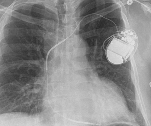
Extensive conduction system abnormalities can have various causes (ischemia, genetic, infectious, amyloid, etc). She was given CRT-D (Cardiac Resynchronization Therapy-Defibrillator). Discussion : The initial ECG in today's case is pathological for any patient, especially for a 50-year old previously heathy female.
She was never defibrillated. 3 of the 4 have similarly bizarre PVCs. == MY Comment by K EN G RAUER, MD ( 4/29/2020 ): == Cardiac Arrest with Bizarre PVCs/Torsades de Pointes: Intriguing case with many interesting features. As was seen in this case — defibrillation and/or overdrive pacing may be needed. Acute ischemia?
12 minutes later, the patient went back into VFib arrest and underwent another 15 minutes of resuscitation followed by successful defibrillation and sustained ROSC. In total, he received approximately 40 minutes of CPR and 7 defibrillation attempts. EMS found the patient in VFib and performed ACLS for 26 minutes then obtained ROSC.
ADDENDUM ( 10/24/2020 ): In the past, the diagnosis of Brugada Syndrome required not only the presence of a Brugada-1 ECG pattern — but also a history of sudden death, sustained VT, non-vasovagal syncope or a positive family history of sudden death at an early age. Smith on this blog ( Simply search for Brugada Syndrome! ).
She was defibrillated and resuscitated. For review of ECG findings expected with Takotsubo Cardiomyopathy — Please see My Comment at the bottom of the page in the March 25, 2020 post in Dr. Smith's ECG Blog ). Upon arrival to the emergency department, a senior emergency physician looked at the ECG and said "Nothing too exciting."
Defibrillation was performed, and ROSC was achieved. The other challenge posed by the ECG of a patient with marked LVH with "strain" is distinguishing between the ST-T wave inversion in one or more lateral leads due solely to LVH vs that due to acute ischemia or infarction.
We organize all of the trending information in your field so you don't have to. Join thousands of users and stay up to date on the latest articles your peers are reading.
You know about us, now we want to get to know you!
Let's personalize your content
Let's get even more personalized
We recognize your account from another site in our network, please click 'Send Email' below to continue with verifying your account and setting a password.


Let's personalize your content