This site uses cookies to improve your experience. To help us insure we adhere to various privacy regulations, please select your country/region of residence. If you do not select a country, we will assume you are from the United States. Select your Cookie Settings or view our Privacy Policy and Terms of Use.
Cookie Settings
Cookies and similar technologies are used on this website for proper function of the website, for tracking performance analytics and for marketing purposes. We and some of our third-party providers may use cookie data for various purposes. Please review the cookie settings below and choose your preference.
Used for the proper function of the website
Used for monitoring website traffic and interactions
Cookie Settings
Cookies and similar technologies are used on this website for proper function of the website, for tracking performance analytics and for marketing purposes. We and some of our third-party providers may use cookie data for various purposes. Please review the cookie settings below and choose your preference.
Strictly Necessary: Used for the proper function of the website
Performance/Analytics: Used for monitoring website traffic and interactions
Written by Jesse McLaren Two patients in their 70s presented to the ED with chestpain and RBBB. Patient 1 : a 75 year old called paramedics with one day of left shoulder pain which migrated to the central chest, which was worse with deep breaths. Ten days later the patient returned with worsening pleuritic chest.
Smith interpretation: This is highly likely to be due to extreme right heart strain and is nearly diagnostic of pulmonary embolism. She had been sitting doing work when she experienced "waves of chest tightness". She had been sitting doing work when she experienced "waves of chest tightness". It is of course pulmonary embolism.
The patient's nitro was dialed up to 100 mcg/min but the pain persisted. The ACC/AHA guidelines mandate less than 2 hours cath for patients with ACS with refractory pain, pulmonary edema, or electrical or hemodynamic instability. Figure-1: I've labeled the initial ECG in today's case ( See text ).
No prior exertional complaints of chestpain, dizziness, lightheadedness, or undue shortness of breath. He denied headache or neck pain associated with exertion. I sent this ECG to Dr. Smith, with the only information that it is a 17 year old with chestpain. 24 yo woman with chestpain: Is this STEMI?
KEY Point: Although true that patients with longstanding, severe pulmonary disease may manifest a QRST complex in standard lead I with marked overall reduction in QRST amplitude ( See ECG Blog #65 — regarding Schamroth’s Sign ) — you should never normally see a completely flat line in any of the standard limb leads.
Written by Pendell Meyers, with some edits by Smith A man in his 40s with many comorbidities presented to the ED with chestpain, hypotension, dyspnea, and hypoxemia. The bedside echo showed a large RV (Does this mean there is a pulmonary embolism as the etiology?) An 80-something woman who presented with chestpain and dyspnea.
An 80-something woman who presented with chestpain and dyspnea. An old formal echo was found from 6 mo ago: Dilated right ventricle with septal flattening and estimated right ventricular systolic pressure of 70 mmHg (significant pulmonary hypertension). After all, this patient did also present with chestpain. ) — See below.
No chestpain. In the context of today's case — these P waves are diagnostic of RAE = P Pulmonale ( See ECG Blog #75 ) and almost certain associated pulmonary hypertension. Working through a case of a regular WCT Rhythm in this 80-something woman — See My Comment in the May 5, 2020 post on Dr. Smith’s ECG Blog.
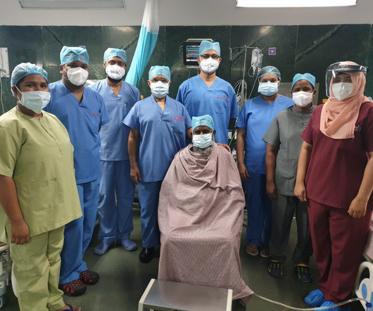
Prateek Bhatnagar, Director Cardiac Surgery & Chief Cardiology Surgeon at Care Hospital, Banjara Hills, Hyderabad, has successfully operated & discharged India’s first post Covid 19 recovered patient who on 16th July, 2020, underwent a triple bypass surgery under Dr. Bhatnagar & his team of doctors here.
I've previously discussed the interesting correlation of a qR pattern in lead V1 in patients with RVH — as strongly suggesting associated pulmonary hypertension ( See ECG Blog #234 and Blog #248 ). This explains the marked increased in QRS amplitude in the lateral chest leads — as well as some of the ST-T wave depression in these leads.
My answer: "This is classic for PE, but it can also be present in any hypoxia due pulmonary hypoxic vasoconstriction and resulting acute pulmonary hypertension and acute right heart strain. An ECG was texted to me (Smith) without any clinical information: What did I say? This is NOT Wellens. Is the patient hypoxic? The answer was yes.
He said that his pain does not feel like his previous episode of pericarditis, and is not related to meals. He denied chestpain, shortness of breath, nausea, fever, chills, rashes, cough, and leg pain. Does subsegmental pulmonary embolism matter? The ST/T ratio in V6, however, is slightly greater.
Written by Pendell Meyers, edits by Smith Two patients presented with acute chestpain/pressure. Chest x-ray was read as normal. CT pulmonary angiogram was negative for pulmonary embolism. Two patients with chestpain. In a patient with chestpain — this is simply not a "normal" ST-T wave in lead V2.
There was some dyspnea but no chestpain. Further ultrasound showed no B-lines (no pulmonary edema). 23/WCC — 2/21/2020 ). 23/WCC — 2/21/2020 ). 23/WCC — 2/21/2020 ). 23/WCC — 2/21/2020 ). A young man presented with continuous prolonged generalized weakness, lightheadedness, and presyncope.
There was no chestpain. This suggests that there is pulmonary hypertension and thus possibly RVH. The estimated pulmonary artery systolic pressure is 31 mmHg + RA pressure. Acute posterior OMI would be a prime concern for the ECG in Figure-1 — IF the patient presented with cardiac-sounding chestpain.
This 60-something with h/o COPD and HFrEF (EF 25%) presented with SOB and chestpain. of all cases, and 62% of Veritas® misdiagnoses). == MY Comment , by K EN G RAUER, MD ( 1/5/2020 ): == This case illustrates a number of important teaching points. The patient in this case presented with dyspnea and chestpain.
He denied chestpain or dyspnea throughout. IF you missed the KEY Findings on the pre-hospital ECG of todays case Please take another look at My Comment at the bottom of the page of that February 6, 2020 post. No previous study for comparison. Clinical Course: - He had no events on cardiac monitoring overnight. -
Given her reported chestpain, shortness of breath, and syncope, an ECG was quickly obtained: What do you think? Figure-2: Causes of Low Voltage on ECG ( Figure reproduced from My Comment at the bottom of the page in the November 12, 2020 post in Dr. Smith's ECG Blog ). What is ELECTRICAL ALTERNANS?
Scenario 1 : The patient presents with 24 hours of substernal chestpain. Ninety percent of patients with reperfusion attained a maximum T wave negativity of 3 mm or more within 48 hours after the onset of chestpain in the lead that initially displayed the greatest ST segment elevation. Below is his presentation ECG.
With OMI, all you know is that your patient has some nonspecific chestpain, SOB, shoulder pain etc. So, given that this patient presents with an acute infectious respiratory illness IF she is hemodynamically stable, it would be reasonable to continue to treat her acute pulmonary illness as we look further at this ECG.
We organize all of the trending information in your field so you don't have to. Join thousands of users and stay up to date on the latest articles your peers are reading.
You know about us, now we want to get to know you!
Let's personalize your content
Let's get even more personalized
We recognize your account from another site in our network, please click 'Send Email' below to continue with verifying your account and setting a password.


Let's personalize your content