This site uses cookies to improve your experience. To help us insure we adhere to various privacy regulations, please select your country/region of residence. If you do not select a country, we will assume you are from the United States. Select your Cookie Settings or view our Privacy Policy and Terms of Use.
Cookie Settings
Cookies and similar technologies are used on this website for proper function of the website, for tracking performance analytics and for marketing purposes. We and some of our third-party providers may use cookie data for various purposes. Please review the cookie settings below and choose your preference.
Used for the proper function of the website
Used for monitoring website traffic and interactions
Cookie Settings
Cookies and similar technologies are used on this website for proper function of the website, for tracking performance analytics and for marketing purposes. We and some of our third-party providers may use cookie data for various purposes. Please review the cookie settings below and choose your preference.
Strictly Necessary: Used for the proper function of the website
Performance/Analytics: Used for monitoring website traffic and interactions
The rhythm is sinus bradycardia at a rate just over 50/minute. The February 11, 2020 post ( LA-RA reversal ). The March 18, 2020 post ( LA-RA reversal ). The July 28, 2020 post ( RA-LL reversal ). The August 28, 2020 post ( LA-LL reversal ). The November 19, 2020 post ( LA-LL reversal ).
Resuscitation was initiated and this ECG was obtained: Likely AFib (irregularly irregular) with bradycardia. Consider thrombolytics for OMI when PCI is not an option. == MY Comment by K EN G RAUER, MD ( 1/19/2020 ): == There is a LOT to talk about regarding the series of tracings in this unfortunate case.
There’s competing sinus bradycardia and junctional rhythm, with otherwise normal conduction, borderline right axis, normal R wave progression and voltages. As Smith and Meyers explained in a 2020 article in EM News : “What should we do in the meantime while we are still stuck in the STEMI paradigm in daily practice? What do you think?
IMPRESSION: The finding of sinus bradycardia with 1st-degree AV block + marked sinus arrhythmia + the change in PR interval from beat #5-to-beat #6 — suggests a form of vagotonic block ( See My Comment in the October 9, 2020 post in Dr. Smith's ECG Blog ). Initial high sensitivity troponin I returned at 6ng/L (normal 0.20
Altered Mental Status, Bradycardia == MY Comment , by K EN G RAUER, MD ( 2/2 /2024 ): == Dr. Meyers began today’s case with the clinical challenge of asking you to identify the underlying cause of ECG #2. -- Read this ECG -- Osborn Waves and Hypothermia (this is the "Figure" above) What does LBBB look like in severe hypothermia?
Looking first at the long-lead II rhythm strip — there is significant bradycardia , with a heart R ate just under 40/minute. But the point to emphasize — is that it should only take seconds to recognize that there is bradycardia from significant AV block. = Would you approve her for a nonemergent surgical procedure?
I will leave more detailed rhythm discussion to the illustrious Dr. Ken Grauer below, but this use of calipers shows that the rhythm interpretation is: Sinus bradycardia with a competing (most likely junctional) rhythm. preceding each of the fascicular beats — indicating a faster rate for the escape rhythm compared to the sinus bradycardia ).
Here is his ED ECG: There is bradycardia with a junctional escape. Case continued A bedside ultrasound showed diminished LV EF and of course bradycardia. The February 11, 2020 post ( LA-RA reversal ). The March 18, 2020 post ( LA-RA reversal ). The August 28, 2020 post ( LA-LL reversal ).
Methods We retrospectively screened 2009 patients who underwent pacemaker implantation from 2010 to 2020 in seven institutions. This study aimed to investigate the association between paced QRS mimicking a complete left bundle branch block (CLBBB) and PICM development.
The August 17, 2020 post by me in Dr. Smith's ECG Blog — in which I review the phenomenon of Bradycardia-dependent BBB ( sometimes called "Phase 4" or "paradoxical" block ). ECG Blog #220 — reviews my L IST # 1 : Causes of a regular WCT ( and how to assess hemodynamic stability ). ECG Blog #242 — Reviews rate -related BBB.
The computer called "Sinus Bradycardia" only (implying that everything else is normal. The overreading Cardiologist called it only "Sinus Bradycardia" with no other findings. The rhythm in Figure-1 is sinus bradycardia and arrhythmia. Here is the old ECG from 6 years prior: Notice the inferior T-waves have normal size here.
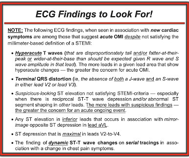
I see the following: The rhythm is sinus bradycardia at ~55-60/minute. The importance of the new OMI ( vs the old STEMI ) Paradigm — See My Comment in the July 31, 2020 post in Dr. Smith's ECG Blog. That said, even without more history and without a prior ECG for comparison — the initial ECG in Figure-1 clearly is of concern!
After the heart rate increased slightly, here was the repeat ECG: Sinus bradycardia, only slightly faster rate than prior. As emphasized in My Comment at the bottom of the page of the June 3, 2020 post in Dr. Smith's ECG Blog — Almost everyone gets fooled the 1st time they see this phenomenon.
For example — marked bradycardia with unusual forms of advanced AV block — or marked bradycardia without evident P waves — or marked QRS widening with "shapeless" morphology — are all ECG indication of the need for immediate IV calcium until improvement of these ECG patterns. Why Isn't the Serum K+ Higher than 6.2
I have periodically called attention to examples of the Ashman phenomenon as they occur in Dr. Smith's ECG Blog ( See My Comments in the January 5, 2020 post — the June 17, 2020 post — and the March 30, 2023 post , among others ).
He had multiple episodes of bradycardia and nonsustained ventricular tachycardia. Angiography revealed a 30% nonobstructive stenosis of the mid LAD. Serial high sensitivity troponin T (URL 15 ng/L) values were negative and stagnant. Patient 1 remained in the hospital overnight. He went to the cath lab at 0900 the next morning.
There is sinus bradycardia with one PVC. Shark Fin morphology has been discussed a number of times on Dr. Smith’s ECG Blog ( For review — See the June 11, 2018 post and the January 24, 2020 post , to name just 2 instances ). 24, 2020 post ( link to that post given above ). She then had a 12-lead: What do you think?
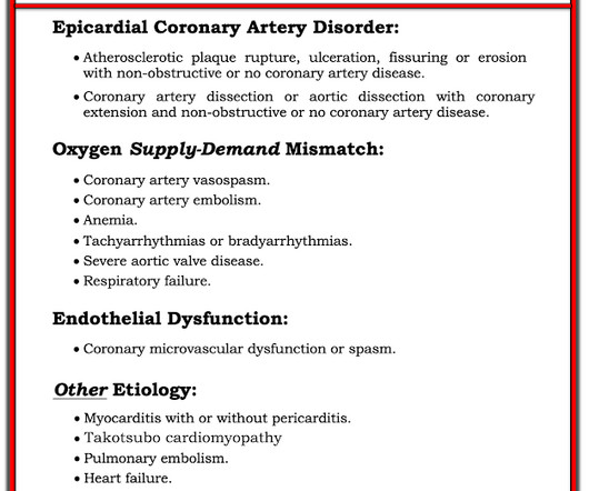
There are 3 etiologies I always think of with bradycardia and AV block: 1. See this terrible case: Computer often fails to diagnose atrial fibrillation in ventricular paced rhythm, and that can be catastrophic == MY Comment , by K EN G RAUER, MD ( 1/22/2020 ): == Our THANKS to Dr. Smith for presenting this extremely interesting case.
The patient later settled into sinus bradycardia. He was started on amiodarone and had no more events. Next day, the cardiologists were convinced (I think correctly) that this was SVT with aberrancy that was triggered by DKA.
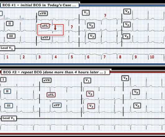
The Initial ECG in Today's Case: ECG #1 showed sinus bradycardia at a rate slightly under 60/minute — normal intervals — slight left axis ( about -15 degrees ) — and no chamber enlargement. Figure-1: I've labeled the 1st, 2nd and 4th tracings in today's case ( See text ).
Sinus bradycardia, normal conduction, normal axis, normal R wave progression, no hypertrophy. JAMA Cardiol 2020 5. -- Litell JM, Meyers HP, Smith SW. The September 21 , 2020 post in Dr. Smith's ECG Blog — My Comment ( at the bottom of the page ) emphasizes utility of the Mirror Test for diagnosis of acute Posterior MI.
Whatever today's rhythm turns out to be — the "good news" is that the bradycardia and degree of AV block is likely to improve as soon as there is reperfusion of the "culprit" artery ( Therefore need for prompt cath with PCI ). For my clarifying Figure illustrating T-QRS-D ( 2nd bullet ) — See My Comment at the bottom of the page in Dr.
3 of the 4 have similarly bizarre PVCs. == MY Comment by K EN G RAUER, MD ( 4/29/2020 ): == Cardiac Arrest with Bizarre PVCs/Torsades de Pointes: Intriguing case with many interesting features. . _ Below, I post 4 more examples of ECGs that were recorded from patients who had Torsades, either shortly before, during, or after.
Her vital signs were within normal limits except for bradycardia at 55 bpm. It is probably sinus bradycardia with very small/depressed P-waves and prolonged PR interval. P EARL # 4 In my opinion, it is not worth wasting time trying to figure out the specific rhythm diagnosis of a bradycardia when there is hyperkalemia.
Figure-1: Reasons for the varied ECG presentation of acute LMain occlusion — excerpted from Dr. Smith’s 8/9/2019 post ( This Table from My Comment in the January 16, 2020 post ). As per Dr. Nossen — today's initial ECG ( LEFT tracing in Figure-2 ) shows sinus bradycardia with QRS widening due to bifascicular block ( RBBB/LAHB ).
There was no evidence bradycardia leading up to the runs of PMVT ( as tends to occur with Torsades ). If there had been — a temporary atrial pacemaker could have been considered as a way of increasing the heart rate to suppress a bradycardia-dependent arrhythmia ("overdrive pacing").
For example — bradycardia and AV conduction disturbances are not uncommon with Hyperkalemia , with these conduction disturbances most often resolving once serum K+ is corrected. Figure-3: Diagnostic considerations for a patient who presents in AV block ( adapted from Mangi et al — StatPearls, 2021 ).
plaque disruption), the T waves still manifest markings of a previous state of suboptimal coronary flow that resolved: Type II supply-demand mismatch in the setting of extreme bradycardia. 2] Although the clinical context in today’s case does not fit these descriptors for Type I OMI (e.g. Phase IV block, or concealed transeptal conduction).
Hyperkalemia causes peaked T waves and the "killer B's of hyperkalemia", including bradycardia, broad QRS complexes, blocks of the AV node and bundle branches, Brugada morphology, and otherwise bizarre morphology including sine wave. With a twist. Do you recognize this ECG yet? Right Bundle Branch Block with ST Elevation in V1?
And a complication. == MY Comment by K EN G RAUER, MD ( 10/11/2020 ): == It cannot be stated any clearer than what Dr. Meyers states above. PEARL #3: I’ve described the Mirror Test on a number of occasions in Dr. Smith’s ECG Blog ( Please see My Comment in the September 21, 2020 post). "It isn't a STEMI," so cath lab refusal (again).
ADDENDUM ( 10/24/2020 ): In the past, the diagnosis of Brugada Syndrome required not only the presence of a Brugada-1 ECG pattern — but also a history of sudden death, sustained VT, non-vasovagal syncope or a positive family history of sudden death at an early age. Smith on this blog ( Simply search for Brugada Syndrome! ).
Theres sinus bradycardia, borderline PR interval, narrow QRS; normal axis/R wave progression; low precordial voltages, and subtle peaked T waves (most obvious in V2, but all T waves are symmetric with a narrow base). Theres no prior ECG to compare - but the bradycardia, prolonged PR and peaked T waves could all be from hyperkalemia.
There is also STE in V1 which is diagnostic of right ventricular OMI in this situation , and partly explains the syncope and hypotension (along with the bradycardia). For more on the ECG diagnosis and consequences of acute RV MI Check out My Comment in July 19, 2020 post and the August 2, 2024 post). This is not the case.
Despite the baseline artifact theres sinus bradycardia, convex ST elevation in III, reciprocal ST depression in aVL and possible anterior ST depression indicating inferoposterior OMI. Still, an ECG was obtained at 0649hrs: (Digitized by PM Cardio) McLaren: The patient has a high pretest probability based on age, risk factors and symptoms.
A repeat ECG was performed as adult cardiology was asked to evaluate the patient for emerget PCI: Sinus bradycardia with persistent elevation in the inferior leads with reciprocal depression in aVL Patient was taken to cath lab with adult cardiology which revealed normal coronary arteries without evidence of occlusion MI.
A post-cath EKG was recorded at 0719: The computer interpretation read Sinus bradycardia, otherwise normal ECG. html ) Despite an undetectable troponin and three normal EKGs, the nature of the patients symptoms and his positive cardiac history warranted concern for ACS. The troponin peaked at 0.4 ng/mL, which is relatively low for OMI.
This ECG shows a sinus bradycardia with a normal conduction pattern (normal PR, normal QRS, and normal QTc), normal axis, normal R-wave progression, normal voltages. Hypothermia can also produce bradycardia and J waves, with a pseudo-STEMI pattern. Internat J Arrhyth 2020 Uesako H, Fukikawa H, Hashimoto S, et al. As per Drs.
We organize all of the trending information in your field so you don't have to. Join thousands of users and stay up to date on the latest articles your peers are reading.
You know about us, now we want to get to know you!
Let's personalize your content
Let's get even more personalized
We recognize your account from another site in our network, please click 'Send Email' below to continue with verifying your account and setting a password.


Let's personalize your content