This site uses cookies to improve your experience. To help us insure we adhere to various privacy regulations, please select your country/region of residence. If you do not select a country, we will assume you are from the United States. Select your Cookie Settings or view our Privacy Policy and Terms of Use.
Cookie Settings
Cookies and similar technologies are used on this website for proper function of the website, for tracking performance analytics and for marketing purposes. We and some of our third-party providers may use cookie data for various purposes. Please review the cookie settings below and choose your preference.
Used for the proper function of the website
Used for monitoring website traffic and interactions
Cookie Settings
Cookies and similar technologies are used on this website for proper function of the website, for tracking performance analytics and for marketing purposes. We and some of our third-party providers may use cookie data for various purposes. Please review the cookie settings below and choose your preference.
Strictly Necessary: Used for the proper function of the website
Performance/Analytics: Used for monitoring website traffic and interactions
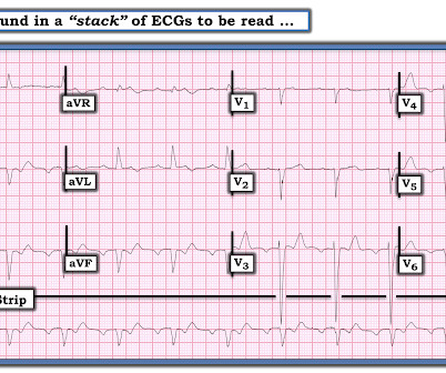
Written by Pendell Meyers, with some edits by Smith A man in his 40s with many comorbidities presented to the ED with chestpain, hypotension, dyspnea, and hypoxemia. The rhythm is 2:1 atrialflutter. An 80-something woman who presented with chestpain and dyspnea. Here is his triage ECG: What do you think?
That volatile course included Atrialflutter with RVR: == MY Comment , by K EN G RAUER, MD ( 7/11 /2023 ): == It's always rewarding to get "a Save!" — as in today's case, in which this 40-something year old patient with persistent VFib, followed by an extended complicated course — ultimately survived with intact neurologic status!
To me, it was clearly atrialflutter with 1:1 conduction. The rate of 280 is just right for atrialflutter. The waves look like atrialflutter waves, NOT like a wide ventricular complex. Recently diagnosed with intermittent paroxysmal atrial fibrillation but no EKGs available to confirm.
She presented to the emergency department after a couple of days of chest discomfort. The ECG was interpreted as showing atrialflutter with 2:1 conduction. The heart rate could be compatible with that of a 2:1 conducted atrialflutter. Also, lead I could give the initial impression of showing flutter waves.
So this is an extremely slow atrialflutter with 2:1 conduction. Atrial rate 146, ventricular rate 73. I suspect that the amyloid slows the conduction of the atrialflutter. It turned out that he had a history of slow atrialflutter. There was no chestpain — and all troponins were negative.
She reports that she is now unable to vagal out of her palpitations and is having shortness of breath and dull chestpain. The differential of a regular narrow QRS tachycardia is sinus tachycardia, SVT, and atrialflutter with regular conduction. Her initial EKG is below.
This 60-something with h/o COPD and HFrEF (EF 25%) presented with SOB and chestpain. of all cases, and 62% of Veritas® misdiagnoses). == MY Comment , by K EN G RAUER, MD ( 1/5/2020 ): == This case illustrates a number of important teaching points. The patient in this case presented with dyspnea and chestpain.
This middle-aged man with no cardiac history but with significant history of methamphetamin and alcohol use presented with chestpain and SOB, worsening over days, with orthopnea. There is atrial activity before every QRS, but that activity has negative polarity, so it is not sinus rhythm. The other atrialflutter types are: 1.
Possible but, again, the QRS morphology is atypical 3) AtrialFlutter with 2:1 conduction and "aberrancy". I do not see flutter wave baseline, and again the QRS morphology is not typical for a supraventricular rhythm. With OMI, all you know is that your patient has some nonspecific chestpain, SOB, shoulder pain etc.
We organize all of the trending information in your field so you don't have to. Join thousands of users and stay up to date on the latest articles your peers are reading.
You know about us, now we want to get to know you!
Let's personalize your content
Let's get even more personalized
We recognize your account from another site in our network, please click 'Send Email' below to continue with verifying your account and setting a password.


Let's personalize your content