This site uses cookies to improve your experience. To help us insure we adhere to various privacy regulations, please select your country/region of residence. If you do not select a country, we will assume you are from the United States. Select your Cookie Settings or view our Privacy Policy and Terms of Use.
Cookie Settings
Cookies and similar technologies are used on this website for proper function of the website, for tracking performance analytics and for marketing purposes. We and some of our third-party providers may use cookie data for various purposes. Please review the cookie settings below and choose your preference.
Used for the proper function of the website
Used for monitoring website traffic and interactions
Cookie Settings
Cookies and similar technologies are used on this website for proper function of the website, for tracking performance analytics and for marketing purposes. We and some of our third-party providers may use cookie data for various purposes. Please review the cookie settings below and choose your preference.
Strictly Necessary: Used for the proper function of the website
Performance/Analytics: Used for monitoring website traffic and interactions
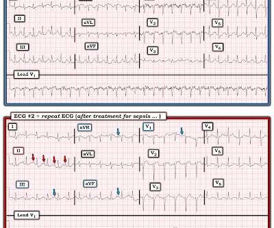
A fully upright P-wave is typical atrial activity of atrialflutter as seen in V1. See these example cases of upright P-waves: Case Continued Thus, I was all but certain that this was atrialflutter. I'd add the following thoughts to Dr. Smith's discussion.
To me, it was clearly atrialflutter with 1:1 conduction. The rate of 280 is just right for atrialflutter. The waves look like atrialflutter waves, NOT like a wide ventricular complex. Reverted to atrial fibrillation with RVR while in the hospital 3 times and needed cardioversion.
This narrows our differential for the rhythm down to sinus tachycardia, paroxysmal supraventricular tachycardia (PSVT, or SVT), and atrialflutter. The patient’s history is notable for paroxysmal atrial fibrillation, which raises clinical suspicion for atrialflutter, since these two entities frequently coexist on a spectrum.
ii ) A reentry SVT ( either AVNRT if the reentry circuit is contained within the AV node — or AVRT if an accessory pathway outside the AV node is involved ) ; iii ) Atrial Tachycardia ( ATach ) ; iv ) AtrialFlutter ( AFlutter ) with 2:1 AV conduction. ECG Blog #138 — AFlutter vs Atrial Tachycardia. s in Figure-2 ).
The ECG was interpreted as showing atrialflutter with 2:1 conduction. The heart rate could be compatible with that of a 2:1 conducted atrialflutter. Also, lead I could give the initial impression of showing flutter waves. She presented to the emergency department after a couple of days of chest discomfort.
PubMed was queried for entries on AF and rurality: (atrial fibrillation OR atrialflutter) AND (rural OR urban OR rurality OR metro OR metropolitan) AND (united states OR US OR U.S.) These studies ranged from 1993 to 2020 and analyzed approximately 41.7 published up to September 24, 2023. million AF patient encounters.
That volatile course included Atrialflutter with RVR: == MY Comment , by K EN G RAUER, MD ( 7/11 /2023 ): == It's always rewarding to get "a Save!" — as in today's case, in which this 40-something year old patient with persistent VFib, followed by an extended complicated course — ultimately survived with intact neurologic status!
PEARL # 3: AtrialFlutter with 1:1 AV conduction is rare! Since the rate of atrial activity with flutter in adults is most often very close to 300 /minute ( ie, usual range for atrial activity ~250-350/minute ) — AFlutter with 2:1 AV conduction typically results in a regular ventricular rate of ~140-160/minute.
The August 17, 2020 post by me in Dr. Smith's ECG Blog — in which I review the phenomenon of Bradycardia-dependent BBB ( sometimes called "Phase 4" or "paradoxical" block ). ECG Blog #220 — reviews my L IST # 1 : Causes of a regular WCT ( and how to assess hemodynamic stability ). ECG Blog #242 — Reviews rate -related BBB.
So this is an extremely slow atrialflutter with 2:1 conduction. Atrial rate 146, ventricular rate 73. I suspect that the amyloid slows the conduction of the atrialflutter. It turned out that he had a history of slow atrialflutter. It turns out that this patient has cardiac amyloidosis.
The principal differential diagnosis of a regular SVT rhythm includes 4 entities: i ) Sinus tachycardia; ii ) A reentry SVT ( either AVNRT if the reentry circuit is contained within the AV node — or AVRT if an accessory pathway located outside the AV node is involved ) ; iii ) Atrial Tachycardia; or , iv ) AtrialFlutter with 2:1 AV conduction.
The differential of a regular narrow QRS tachycardia is sinus tachycardia, SVT, and atrialflutter with regular conduction. There are no P waves preceding the QRS complexes, and no clear flutter waves. We see a regular tachycardia with a narrow QRS complex and no evidence of OMI or subendocardial ischemia.
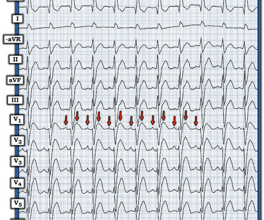
The rhythm is 2:1 atrialflutter. The flutter waves can conceal or mimic ischemic repolarization findings, but here I don't see any obvious findings of OMI or subendocardial ischemia. The bedside echo showed a large RV (Does this mean there is a pulmonary embolism as the etiology?) Here is his triage ECG: What do you think?
of all cases, and 62% of Veritas® misdiagnoses). == MY Comment , by K EN G RAUER, MD ( 1/5/2020 ): == This case illustrates a number of important teaching points. M Y A NSWER: In my experience, MAT is the 2nd-most commonly overlooked cardiac arrhythmia ( surpassed only by AtrialFlutter ).
The Differential Diagnosis is: SVT with aberrancy(#) [AVNRT vs. WPW (also called AVRT*)] Atrialflutter with 1:1 conduction, with aberrancy VT coming from the anterior fascicle ( fascicular VT )@ *AVRT = AV Reciprocating Tachycardia (Tachycardic loop that uses both the AV node and an accessory pathway.
There is atrial activity before every QRS, but that activity has negative polarity, so it is not sinus rhythm. There are clearly no flutter waves, so it is not atrialflutter (a "macro-reentrant" atrial tachycardia) Is it AVNRT originating at the superior pole of the AV node, resulting in a retrograde P-wave before the QRS?
The aim of this study was to examine sex differences in prescription and adherence to GDMT at 1-year after ischemic stroke in a cohort of commercially insured patients.Methods:Using the Truven Health MarketScan database from 2016-2020, we identified patients admitted with ischemic stroke. The mean age was 55.7 vs 45.0%, P=0.003).
Methods and Results Between January 2020 and December 2023, 118 PeAF patients were selected for first intent Marshall plan ablation (MPA). After the blanking period of 3 months, 62/109 patients were in sinus rhythm (SR) (57%), 33/109 were in AF (30.2%), 8/109 were in left atrialflutter (AFL) (7.3%), and six were in right AFL (5.5%).
Note: The ECG in Figure-1 was initially recorded using the Cabrera Format ( See Comment by Dr. Grauer at the bottom of the page in the October 26, 2020 post for review of the Cabrera Format ). Before each of these WCT episodes the atrial rate decreased. Other than sinus rhythm What else do you see in this tracing?
Possible but, again, the QRS morphology is atypical 3) AtrialFlutter with 2:1 conduction and "aberrancy". I do not see flutter wave baseline, and again the QRS morphology is not typical for a supraventricular rhythm. See this case, for example: A Relatively Narrow Complex Tachycardia at a Rate of 180.
We organize all of the trending information in your field so you don't have to. Join thousands of users and stay up to date on the latest articles your peers are reading.
You know about us, now we want to get to know you!
Let's personalize your content
Let's get even more personalized
We recognize your account from another site in our network, please click 'Send Email' below to continue with verifying your account and setting a password.


Let's personalize your content