This site uses cookies to improve your experience. To help us insure we adhere to various privacy regulations, please select your country/region of residence. If you do not select a country, we will assume you are from the United States. Select your Cookie Settings or view our Privacy Policy and Terms of Use.
Cookie Settings
Cookies and similar technologies are used on this website for proper function of the website, for tracking performance analytics and for marketing purposes. We and some of our third-party providers may use cookie data for various purposes. Please review the cookie settings below and choose your preference.
Used for the proper function of the website
Used for monitoring website traffic and interactions
Cookie Settings
Cookies and similar technologies are used on this website for proper function of the website, for tracking performance analytics and for marketing purposes. We and some of our third-party providers may use cookie data for various purposes. Please review the cookie settings below and choose your preference.
Strictly Necessary: Used for the proper function of the website
Performance/Analytics: Used for monitoring website traffic and interactions
A fully upright P-wave is typical atrial activity of atrialflutter as seen in V1. See these example cases of upright P-waves: Case Continued Thus, I was all but certain that this was atrialflutter. If it is flutter, it will reveal the underlying flutter waves. BP was 100 systolic.
Atrialfibrillation (AF) is the most common sustained arrhythmia and associated with increased morbidity and mortality. PubMed was queried for entries on AF and rurality: (atrialfibrillation OR atrialflutter) AND (rural OR urban OR rurality OR metro OR metropolitan) AND (united states OR US OR U.S.)
Here is the computer interpretation: ATRIALFIBRILLATION WITH RAPID VENTRICULAR RESPONSE WITH ABERRANT CONDUCTION OR VENTRICULAR PREMATURE COMPLEXES LEFT AXIS DEVIATION [QRS AXIS beyone -30] NONSPECIFIC ST and T-WAVE ABNORMALITY The over-reading physician confirmed this diagnosis, which is incorrect. It is not atrialfibrillation.
To me, it was clearly atrialflutter with 1:1 conduction. The rate of 280 is just right for atrialflutter. The waves look like atrialflutter waves, NOT like a wide ventricular complex. Recently diagnosed with intermittent paroxysmal atrialfibrillation but no EKGs available to confirm.
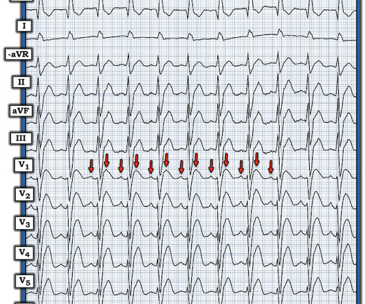
edits by Meyers A woman in her 60s with a history of chronic atrialfibrillation on Eliquis, ESRD on hemodialysis, type-II diabetes mellitus, prior CVA, hypertension, and hyperlipidemia presented to the emergency department with multiple complaints after missing dialysis. They are flutter waves, and the rhythm is 2:1 atrialflutter.
She also has a hx of paroxysmal atrialfibrillation and is on oral anticoagulant treatment. The ECG was interpreted as showing atrialflutter with 2:1 conduction. The heart rate could be compatible with that of a 2:1 conducted atrialflutter. The last echocardiography 12 months ago showed HFmrEF.
The differential of a regular narrow QRS tachycardia is sinus tachycardia, SVT, and atrialflutter with regular conduction. There are no P waves preceding the QRS complexes, and no clear flutter waves. This includes sinus tachycardia, atrialfibrillation or flutter, MAT, and others.
There is atrial activity before every QRS, but that activity has negative polarity, so it is not sinus rhythm. There are clearly no flutter waves, so it is not atrialflutter (a "macro-reentrant" atrial tachycardia) Is it AVNRT originating at the superior pole of the AV node, resulting in a retrograde P-wave before the QRS?
ABSTRACT Background Different ablation strategies have been developed for persistent atrialfibrillation (PeAF), but early management is still controversial. Methods and Results Between January 2020 and December 2023, 118 PeAF patients were selected for first intent Marshall plan ablation (MPA).
Note: The ECG in Figure-1 was initially recorded using the Cabrera Format ( See Comment by Dr. Grauer at the bottom of the page in the October 26, 2020 post for review of the Cabrera Format ). Figure-2: Colored arrows highlight flutter waves , with 2:1 AV conduction. Before each of these WCT episodes the atrial rate decreased.
We organize all of the trending information in your field so you don't have to. Join thousands of users and stay up to date on the latest articles your peers are reading.
You know about us, now we want to get to know you!
Let's personalize your content
Let's get even more personalized
We recognize your account from another site in our network, please click 'Send Email' below to continue with verifying your account and setting a password.


Let's personalize your content