This site uses cookies to improve your experience. To help us insure we adhere to various privacy regulations, please select your country/region of residence. If you do not select a country, we will assume you are from the United States. Select your Cookie Settings or view our Privacy Policy and Terms of Use.
Cookie Settings
Cookies and similar technologies are used on this website for proper function of the website, for tracking performance analytics and for marketing purposes. We and some of our third-party providers may use cookie data for various purposes. Please review the cookie settings below and choose your preference.
Used for the proper function of the website
Used for monitoring website traffic and interactions
Cookie Settings
Cookies and similar technologies are used on this website for proper function of the website, for tracking performance analytics and for marketing purposes. We and some of our third-party providers may use cookie data for various purposes. Please review the cookie settings below and choose your preference.
Strictly Necessary: Used for the proper function of the website
Performance/Analytics: Used for monitoring website traffic and interactions
MY Thoughts on Today’s CASE: As tempting as it might be to reach for the defibrillator on seeing the ECG shown in Figure-1 — My initial reaction was different. The April 6, 2023 post — excessive baseline artifact misdiagnosed as AFib ( instead of sinus rhythm with AV Wenckebach — as in Figure-4 in this post ). Is this VT?
But artifact is "alive and well" — and learning to recognize it will amaze many of your colleagues ( and may serve to avoid an unnecessary defibrillation or two ). The April 6, 2023 post — excessive baseline artifact misdiagnosed as AFib ( instead of sinus rhythm with AV Wenckebach — as in Figure-4 in this post ).
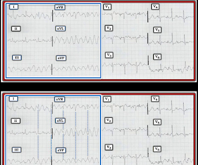
After resuscitation and defibrillation , there were no more episodes of TdP. Below is the patient’s 12 lead ECG following defibrillation. Of note — the QT interval of beat #5 ( blue line ) is markedly prolonged compared to the QT interval in the beginning of the tracing ( red line ). What does this ECG tell you?
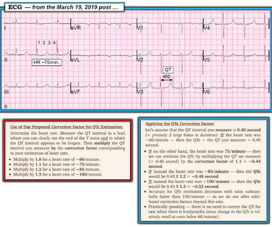
For more on my systematic approach — Check out My Comment in the May 3, 2020 post ). Treatment is by ICD ( implantable cardioverter defibrillator ). Take another LOOK at today's ECG ( which I've reproduced in Figure-1 ): Figure-1: I’ve labeled the initial tracing in today’s case. How Short is the QTc in Today's Tracing?
Rhythm C: This telemetry strip from an older adult was initially thought to need defibrillation. The April 6, 2023 post — excessive baseline artifact misdiagnosed as AFib ( instead of sinus rhythm with AV Wenckebach — as in Figure-4 in this post ). The November 10, 2020 post — for PTA. The March 17, 2023 post — for PTA.
Treatment is by ICD ( implantable cardioverter defibrillator ). Assuming there was no history of cardiac arrest, unexplained syncope or AFib at an early age — cardiac risk from a “short” QTc is clearly less than for patients with frank SQTC. Males with a QTc ≤360 ms — and females with a QTc ≤370 ms are said to have a “ short ” QTc.
We organize all of the trending information in your field so you don't have to. Join thousands of users and stay up to date on the latest articles your peers are reading.
You know about us, now we want to get to know you!
Let's personalize your content
Let's get even more personalized
We recognize your account from another site in our network, please click 'Send Email' below to continue with verifying your account and setting a password.


Let's personalize your content