This site uses cookies to improve your experience. To help us insure we adhere to various privacy regulations, please select your country/region of residence. If you do not select a country, we will assume you are from the United States. Select your Cookie Settings or view our Privacy Policy and Terms of Use.
Cookie Settings
Cookies and similar technologies are used on this website for proper function of the website, for tracking performance analytics and for marketing purposes. We and some of our third-party providers may use cookie data for various purposes. Please review the cookie settings below and choose your preference.
Used for the proper function of the website
Used for monitoring website traffic and interactions
Cookie Settings
Cookies and similar technologies are used on this website for proper function of the website, for tracking performance analytics and for marketing purposes. We and some of our third-party providers may use cookie data for various purposes. Please review the cookie settings below and choose your preference.
Strictly Necessary: Used for the proper function of the website
Performance/Analytics: Used for monitoring website traffic and interactions
The past five years have yielded impressive advancements in fully absorbable metal stent technology. Nowhere is the need for fully absorbable metal stents greater than in patients experiencing vascular anomalies associated with congenital heart disease (CHD).
(Gore) announced recent FDA approval of a lower profile GORE VIABAHN VBX Balloon Expandable Endoprosthesis ( VBX Stent Graft ). 1-3 "Our team is pleased to be the first commercial implanter of the new lower profile VBX Stent Graft," said Darren Schneider , M.D., No changes to the stent design were made to achieve the lower profile.
BACKGROUND:Carotid artery stenting (CAS) is an alternative treatment for patients with carotid artery stenosis who are not eligible for carotid endarterectomy. Stroke, Ahead of Print. Dual antiplatelet therapy (DAPT) after CAS aims to prevent ischemic stroke. However, its optimal duration remains unclear.
To our knowledge, no studies have directly compared the right and left TRA for carotid artery stenting (CAS). The right TRA was performed as a first-line approach from 2019 to 2021, with the left TRA being used thereafter.
Background:Hyperperfusion phenomenon (HPP) constitutes a significant risk factor for adverse outcomes following carotid artery stenting (CAS). Stroke, Volume 56, Issue Suppl_1 , Page ATP178-ATP178, February 1, 2025. Currently, the sole method for evaluating the risk of HPP post-CAS is the invasive acetazolamide (ACZ) challenge test.
It can provide intraluminal fly-through and clipping-plane views which help endovascular assessment of stents, aneurysms, vessel wall irregularities and calcification. 2019 Nov;14(6):1046-1057. 2019 Nov;14(6):1046-1057. Epub 2019 Sep 4. Reference Kang SL, Armstrong A, Krings G, Benson L. Congenit Heart Dis. Cardiol Young.
Unfortunately, we do not have those images for review, but the operators described a ruptured LAD plaque and they stented this area, which ensures the stability of the plaque. The image on the left shows the LAD before intervention, and the red circled portion on the right indicates the stented region.
IntroductionThe 2015 American Heart Association Guidelines recommended mechanical thrombectomy with stent‐retriever devices. We analyze the national trends in mechanical thrombectomy use and outcomes for stroke five years after publication of the US guideline update.MethodsWe analyzed the National Inpatient Sample from 2012‐2019.
This led to immediate cath lab activation — which revealed total occlusion of a large 1st diagonal branch that was stented. == Below is the ECG of Patient #3 — recorded from a 35-year old man with sudden, new-onset CP. In a word — Patient #2 was lucky to have his ECG interpreted by the Queen Of Hearts.
At that visit the patient was found to have an in-stent RCA occlusion. The patient was referred emergently to the cath lab, and again there was an in-stent RCA occlusion. In fact, this patient had an extensive hx of prior MIs. The ECG (ECG#2) on file that was given you was obtained three months prior. Troponin I peaked at 18.323ng/L.
The studies were classified in 3 periods by publication date: Period 1: before 2015, pre-stent retriever; Period 2: 2015-2019, early thrombectomy era; and Period 3: 2020-2024, recent period.Results:Of 2693 references, 21 trials met inclusion criteria, 3 in Period 1, 10 in Period 2, 8 in Period 3.
first reported 3 patients with vertebral webs diagnosed by DSA and having MRI show ischemic strokes in the vascular territory were treated with endovascular stenting and had no recurrence of an ischemic event (4). Lenck et al.
7 The use of antiplatelet agents to prevent stent thrombosis, moderate- to high-dose statin therapy after acute coronary syndromes, or antihypertensive agents in asymptomatic patients may all be perceived by patients as not providing benefit because they may not feel the effects.8
Two adjunctive modifications of CE ‐ balloon remodeling techniques (BRT) and stent‐assisted coiling (SAC) ‐ have been utilized to facilitate occlusion of BTAs of variable anatomies/morphologies, sizes, and rupture status. Stents approved by FDA after 2014 (used in 13 cases) had a greater rate of retreatment (46.2% vs. 10.7%).
We excluded patients who had a clear contraindication to Eptifibatide, received a stent, or if the luminal stenosis was related to reactive vasospasm and any cases with TICI 0, 1, or TICI 3 scores.Results:Our sample size was 60 (51.7% female, mean age 63.9).
A prehospital “STEMI” activation was called on a 75 year old male ( Patient 1 ) with a history of hyperlipidemia and LAD and Cx OMI with stent placement. It was stented. He wrote most of it and I (Smith) edited. Still, such dramatic changes cannot be overlooked. V2 has some features of type 2 Brugada phenocopy. This was a large OMI.
Here we showed the efficacy and safety of PCSK9 inhibitor injection immediately prior to mechanical thrombectomy for these patients.MethodsThe subjects were stroke patients who underwent mechanical thrombectomy at Inha University Hospital from April 2019 to March 2023. vs 53.4%, p = 0.04).
IntroductionTo provide our single‐institution experience and outcome data with the WEB device in 51 patients treated for ruptured and unruptured intracranial aneurysms.MethodsOcclusion rates in a cohort of 51 patients treated with WEB were collected at time of procedure and at last follow‐up between the years 2019 and 2021.
One would not expect wall motion to recover so quickly after stenting, so this is good evidence that the POCUS echo was indeed accurate. Angiogram: Severe diffuse left main disease, up to 80% at the ostial left main. Post cath ECG: Normal or near normal Peak troponin I was 15 ng/mL.
Clinical outcome was treatment‐related visual complications.ResultsA total of 60 patients treated at our tertiary center between January 2017 and November 2019 were reviewed. Imaging outcomes were aneurysm occlusion and OA patency at 1‐year, determined by Digital Subtraction Angiography (DSA) or MRA. 4 patients were lost to follow up.
The patient is female in her 80s with a medical hx of previous MI with PCI and stent placement. the most commonly overlooked arrhythmia ( See My Comment at the bottom of the page in the May 1, 2023 — and the November 12, 2019 post , among others ). The last echocardiography 12 months ago showed HFmrEF.
It was opened and stented with resulting TIMI-3 (normal) flow. The stent to LCX is patent. When I didn’t — I went back to the August 9, 2019 Post that Dr. Smith references in his comments above. I have summarized the major points from Dr. Smith’s 8/9/2019 post in Figure-1. OM1 is occluded and OM2 has 60% stenosis.
5 ICSS‐ MRI study (International Carotid Stenting Study Magnetic Resonance Imaging Study), indicated that patients with periprocedural hemodynamic depression had decreased cerebral blood flow and increased the risk of new lesions in imaging.6 This is secondary to delayed postoperative cerebral ischemia and infarction caused by vasospasm.7
LAD and D1 were stented, but flow unfortunately could not be well restored despite efforts (they list the post intervention TIMI flow still as 0). We wrote this Editorial in the Journal of Electrocardiology in 2019. Serial tracings following stent placement confirmed the large extent of myocardial injury.
60-something with h/o MI and stents presented with chest pain radiating to the back and nausea/vomiting. It was stented. The patient had a p rior h istory of MI + stents. Time zero What do you think? There is inferior ST elevation. Is it normal variant? Is it ischemic (OMI)? Pericarditis? Compare with an old ECG.
Troponin I returned 80 ng/mL, and the Cath Lab was then reactivated where a 100% LAD occlusion was found and stented. The nearest PCI center was activated but ultimately denied by Cardiology. Chapter 22: Electrolytes, Temperature, Central Nervous System Disease, and Miscellaneous Effects. Elsevier-Saunders: Philadelphia, PA. [5]. McCance, K.
They found 100% acute mid-LAD Occlusion MI, stented with excellent angiographic result. Ongoing ischemia (by symptoms, ECG, or troponin) despite maximal medical management is an indication for emergent cath lab activation. == MY Comment by K EN G RAUER, MD ( 8/15/2019 ): == Once again, the w rong q uestion was a sked in this case.
It was a 60yo with a history of stents to the circumflex and right coronary arteries, who presented with 9 hours of fluctuating central chest pain. Int J Cardiol 2019 2. -- Meyers HP, Bracey, Smith et al. J Electrocardiol 2019 6. -- Amsterdam EA, Wenger NK, Brindis RG, et al. Ischemic ST depression maximal in V1-V4 (vs.
A mid-LAD culprit lesion was identified and stented. Cardiology felt her chest pain to be, most likely, the result of coronary supply-demand mismatch in the context of HCM endothelial remodeling (i.e. Type II MI), however decided to pursue coronary angiogram out of an abundance of caution. References Naidu, S. American College of Cardiology.
100% proximal LAD successfully stented. I recognized this as a STEMI immediately and I was only able to do so solely because of your blog." == Comment by K EN G RAUER, MD ( 8/18/2019 ): == Our thanks to Dr. "Cardiology agreed to take the pt to the lab but thought it would likely be negative. Defibrillated out of v fib in the cath lab.
Troponin profile The patient underwent angiography and had a 90% thrombotic proximal LAD lesion that was stented. See this: Classic Evolution of Wellens' T-waves over 26 hours And a bit of further evolution here. The estimated left ventricular ejection fraction is 61 %.
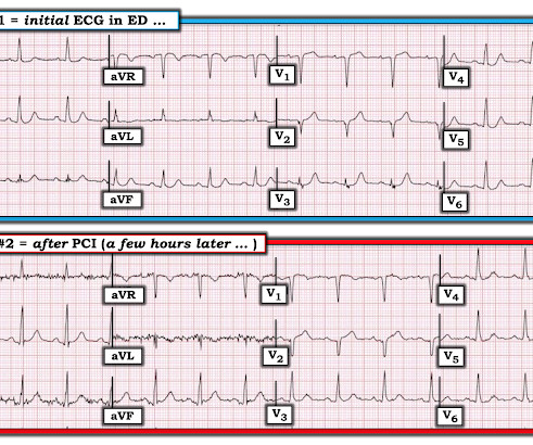
The culprit lesion was opened and stented. For more on this mirror-image opposite ST-T wave relation in leads III vs aVL — See My Comment in the March 8, 2019 and August 9, 2018 posts in Dr. Smith's ECG Blog ). The QoH now recognizes the OMI with mid confidence. Below is the post -PCI electrocardiogram.
It was stented. == MY Comment by K EN G RAUER, MD ( 9/27/2019 ): == As suggested by the title of this Blog post — confirmation of the diagnosis in this case was made not by ECG — but instead by chest aorta CT !
Here is the post stent ECG: This is probably the amount of ST elevation (zero) that this patient has at baseline. At angiogram with a rapid door to balloon time: Culprit is 100% occlusion of the LAD Mid segment. Note the beginning of Wellens' (reperfusion T-waves) in V3-V6.
The 50-something patient with history of coronary stenting and slightly reduced LV ejection fraction. In the setting of prior stenting and reduced left ventricular ejection fraction, would pursue a heart team revascularization approach Syntax score 28.5, This alone could be due to LVH, but V4 could NOT be due to LVH.
They were stented. We wrote this Editorial in the Journal of Electrocardiology in 2019. [link] Unbeknownst to us at the time, there was an old ECG for comparison from 3.5 years prior which I only found a day later: This is a truly normal ECG, with normal sized T-waves and normal S-waves in V2 and V3. The peak troponin was 1863 ng/L.
The lesion was successfully stented, but it was unfortunately done after a significant myocardial loss. 2019 Apr;21(5):253-258. You may see a filling defect in distal LAD, most probably due to an embolization from proximal lesion. Also note that LAD does not extensively wrap-around apex and supply inferior wall. Anatol J Cardiol.
Slow TIMI 2 initially with brisk flow status post percutaneous coronary intervention with 18mm drug-eluting stent. He was taken emergently to the cardiac catheterization lab and found to have multi-vessel coronary artery disease with a near-occlusive culprit lesion in the RCA, possibly reperfused. To our knowledge, the patient did well.
100% proximal LAD thrombotic occlusion with TIMI 0 flow was found and stented with excellent angiographic result and TIMI 3 flow. When in doubt, record serial ECGs and watch out for signs of ischemia despite medical management. == Comment by K EN G RAUER, MD ( 7/11/2019 ): == Our thanks to Drs. Cath images: Before intervention.
It was opened and stented. link] == MY Comment, by K EN G RAUER, MD ( 12/17/2019 ): == Great case about some subtleties in association with LBBB. Chest pain with New LBBB: It helps to actually measure the ST/S ratio A Fascinating Demonstration of ST/S Ratio in LBBB and Resolving LAD Ischemia The cath lab was activated.
Thrombectomy performed, then stent placed with improvement of TIMI 0 to TIMI 3 flow. Always keep in mind the possibility of diffuse STE (and other OMI findings) as being due to a type 3 LAD. == Comment by K EN G RAUER, MD ( 7/19/2019 ): == I was intrigued by the challenge of clinical correlation posed by the ECGs in this case.
I describe and illustrate in detail my approach to incorporating these criteria in My Comment at the bottom of the December 16, 2019 post in Dr. Smiths ECG Blog. There was 100% occlusion of the RCA, which was stented. Dr. Smith illustrates how to measure these parameters with magnified views in his December 21, 2015 post.
They had a history of non-ischemic cardiomyopathy (EF 30%), as well as PCI with one stent. Case contributed by Brooks Walsh , an emergency physician and ECG aficionado in Connecticut (and a few comments by Smith) A middle-aged patient came to the ED complaining of palpitations, dyspnea, sweating, and chest pressure that radiated to the arms.
We organize all of the trending information in your field so you don't have to. Join thousands of users and stay up to date on the latest articles your peers are reading.
You know about us, now we want to get to know you!
Let's personalize your content
Let's get even more personalized
We recognize your account from another site in our network, please click 'Send Email' below to continue with verifying your account and setting a password.


Let's personalize your content