Remove 2019 Remove Electrocardiogram Remove Myocardial Infarction
article thumbnail

Use of downstream stress imaging tests for risk stratification of patients presenting to the emergency department with chest pain and low HEART score

Open Heart

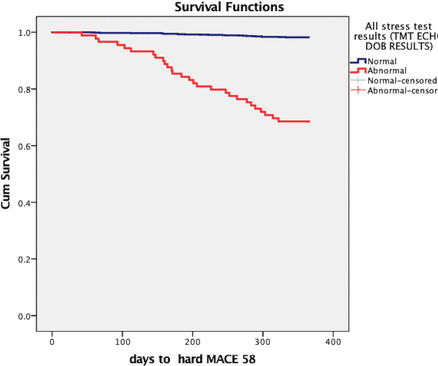
Background Patients with low HEART (History, Electrocardiogram, Age, Risk factors, and Troponin level) risk scores who are discharged from the emergency department (ED) may present clinical challenges and diagnostic dilemmas. Methods We prospectively included 1384 patients with LRHSs between March 2019 and March 2021.

article thumbnail

Cardiovascular disease development in COVID-19 patients admitted to a tertiary medical centre in Iran

The British Journal of Cardiology

Cardiovascular diseases (CVDs) have been reported to occur in a significant number of patients diagnosed with coronavirus disease 2019 (COVID-19). Cardiovascular consultation had been requested for all of the patients based on their primary clinical examination, vital signs, and electrocardiogram (ECG).

article thumbnail

Safety and Effectiveness of the High-Sensitivity Cardiac Troponin HEART Pathway in Patients With Possible Acute Coronary Syndrome

Circulation: Cardiovascular Quality & Outcomes

BACKGROUND:The HEART Pathway (History, Electrocardiogram, Age, Risk factors, Troponin) can be used with high-sensitivity cardiac troponin to risk stratify emergency department patients with possible acute coronary syndrome. Circulation: Cardiovascular Quality and Outcomes, Ahead of Print. The hs-HP was also associated with 16.1% (95% CI, 14.9%–17.3%)

article thumbnail

New ECG Study Reveals Myocarditis Risk in Athletes after Covid-19.

Wellnest

The World Health Organisation (WHO) formally proclaimed COVID-19, the illness spread by a zoonotic SARS-CoV-2, as a pandemic in March 2020, after it had started to spread in late 2019. And, addressing myocardial infarction, recent studies have shown that 1-4% of Athletes untimely die due to myocarditis caused by COVID-19.

article thumbnail

Dark Side of the Moon

EMS 12-Lead

The peak Troponin I confirmed myocardial infarction. (A Posterior myocardial infarction: The dark side of the moon. The delayed action wave in non-ST-elevation myocardial infarction. The EMS crews were correct moving forward with STEMI activation. 1] Driver, B. Netherlands Heart Journal, 15 (1), 16-21. [3]

article thumbnail

OMI-NOMI paradigm established as better than STEMI-NSTEMI with new article

Dr. Smith's ECG Blog

Terminal QRS distortion ( ie, the absence of both a J-wave and an S-wave in either lead V2 or lead V3 ) — SEE My Comment in the November 14, 2019 post for an illustration and description of T-QRS-D. The more leads in a given lead area that show hyperacute changes — the greater the concern for acute OMI.

STEMI 52
article thumbnail

90 year old with acute chest and epigastric pain, and diffuse ST depression with reciprocal STE in aVR: activate the cath lab?

Dr. Smith's ECG Blog

Am J Med 2019, 132(5):622-630. New insights into the use of the 12-lead electrocardiogram for diagnosing acute myocardial infarction in the emergency department. Now there is a paper published in 2019 that proves the point beyond doubt, though makes it clear that this pattern is associated with very high mortality.