This site uses cookies to improve your experience. To help us insure we adhere to various privacy regulations, please select your country/region of residence. If you do not select a country, we will assume you are from the United States. Select your Cookie Settings or view our Privacy Policy and Terms of Use.
Cookie Settings
Cookies and similar technologies are used on this website for proper function of the website, for tracking performance analytics and for marketing purposes. We and some of our third-party providers may use cookie data for various purposes. Please review the cookie settings below and choose your preference.
Used for the proper function of the website
Used for monitoring website traffic and interactions
Cookie Settings
Cookies and similar technologies are used on this website for proper function of the website, for tracking performance analytics and for marketing purposes. We and some of our third-party providers may use cookie data for various purposes. Please review the cookie settings below and choose your preference.
Strictly Necessary: Used for the proper function of the website
Performance/Analytics: Used for monitoring website traffic and interactions
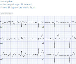
I assumed it was a patient with acute chestpain. It was a man in his 30s with chestpain. This was sent to me from Sam Ghali ( @EM_Resus ) with no other information. What do you think, Steve? Real or just fake?" What do YOU think? It has some inferior ST elevation with some reciprocal ST depression and inverted T in aVL.
By Magnus Nossen This ECG is from a young man with no risk factors for CAD, he presented with chestpain. The patient is a young adult male with chestpain. The chestpain was described as pressure like and radiation to both arms and the jaw. How would you assess this ECG? What is your next step?
Sent by Magnus Nossen MD, written by Pendell Meyers A man in his 50s, previously healthy, developed acute chestpain. The primary care physician there evaluated this patient and deemed the chestpain to be due to gastrointestinal causes. Clinical Cardiology 2019. No troponins were measured!
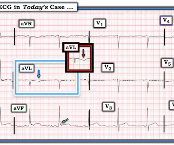
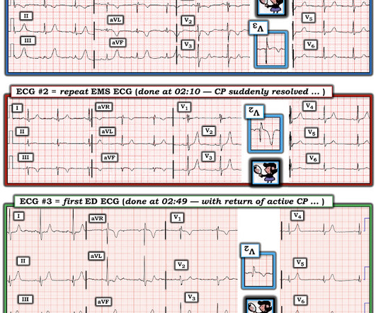
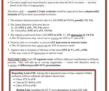
Written by Magnus Nossen with Edits by Grauer and Smith The ECGs in today’s case are from 3 different patients all presenting with new-onset CP ( ChestPain ). We have addresssed this issue on a number of occasions ( See My Comment in the April 25, 2023 — July 21, 2019 — December 10, 2019 — and January 10, 2020 posts).
Written by Jesse McLaren Four patients presented with chestpain. 2019 ; 54 : 79 - 81. All initial ECGs were labeled ‘normal’ or ‘otherwise normal’ by the computer interpretation, and below are the ECGs with the final cardiology interpretation. Am J Emerg Med. 2021 ; 46 : 23 - 26 4. Litell JM , Meyers HP , Smith SW.
Written by Jesse McLaren Two patients in their 70s presented to the ED with chestpain and RBBB. Patient 1 : a 75 year old called paramedics with one day of left shoulder pain which migrated to the central chest, which was worse with deep breaths. Do either, both, or neither have occlusion MI? Vitals were normal.
Written by Pendell Meyers A man in his early 40s experienced acute onset chestpain. The chestpain started about 24 hours ago, but there was no detailed information available about whether his pain had come and gone, or what prompted him to be evaluated 24 hours after onset. Litell JM, Meyers HP, Smith SW.
A healthy 45-year-old female presented with chestpain, with normal vitals. The patient was previously healthy, with no atherosclerotic risk factors, and developed chestpain after an episode of stress. The pain was crushing retrosternal, radiated to the arms and was associated with lightheadedness.
Case An 82 year old man with a history of hypertension presented to the ED with chestpain at 1211. He described his chestpain as pleuritic and reported that it started the day prior while swinging a golf club. His pain suddenly became much worse in the ED and he became acutely diaphoretic, dizzy, and hypotensive.
Healthy male under 25 years old with a pretty good story for acute onset crushing chestpain relieved with nitro. PEARL: Most patients who present with new chestpain + ECG changes + positive troponin — will not need Cardiac MRI. This was sent to me by a partner: "Curious what you think of this one we had overnight.
They had difficulty describing their symptoms, but complained of severe weakness, nausea, vomiting, headache, and chestpain. They described the chestpain as severe, crushing, and non-radiating. Altogether, this strongly suggests inferolateral OMI, particularly in a patient with acute chestpain.
Written by Jesse McLaren A previously healthy 60 year old developed exertional chestpain with diaphoresis, and called EMS. Here’s the EMS ECG, digitized with PM cardio. What do you think? There’s sinus arrhythmia with normal conduction, normal axis and normal voltages. There’s loss of R waves in V2-3 with hyperacute waves V1-5.
[link] A 30 year-old woman was brought to the ED with chestpain. She had given birth a week ago, and she had similar chestpain during her labor. She attributed the chestpain to anxiety and stress, saying "I'm just an anxious person." JACC 2019 Sep 10;74(10):1290-1300.
A previously healthy middle-aged male presented shortly after the acute onset of chestpain very shortly before calling 911. On arrival, he was pain free: What do you think? Jerry Jones commented: "Any ST depression on the ECG of a patient with chestpain credible for ACS represents a reciprocal change until proved otherwise."
male was sitting at a work conference when he began having substernal chestpain with diaphoresis. The pain was 7 out of 10 when this ECG was recorded: The QTc = 375 What do you think? In a patient with worrisome, new-onset chestpain — there is NO way that the T wave in lead V3 is normal.
Sent by anonymous, written by Pendell Meyers A man in his 50s with no prior known medical history presented to the Emergency Department with severe intermittent chestpain. He denied any lightheadedness, shortness of breath, vomiting, or abdominal pain. Isn't it amazing?? Don’t Ignore Bedside Echo Results! —
A 60 year old with chestpain presented to the ED. In this case, lead I does not look bizarre, but all other leads do. == N OTE : The reasons I especially liked today's case are: i ) The patient presented with chestpain — so the importance of distinguishing artifact from reality can not be overstated! —
This is a previously healthy male teenager who was awoken by chestpain. The pain is described as located in the midsternal area, radiating to the right arm, described as 8-9/10 and worse with deep inspirations. In the evening, he became diaphoretic and complained of 9/10 continuous chestpain.
The patient presented with chestpain. I was taught that the tell-tale sign of ischemia vs an electrical abnormality was in the hx, i.e. chestpain for the ischemia and potential syncope for brugada. The patient in question presented with new-onset chestpain — and was from South(east) Asia.
An elderly woman presented with chestpain that radiated to the back for several hours. The first troponin returned at 0.099 ng/mL (elevated, consistent with Non-Occlusion MI) Providers were concerned with aortic dissection, so they order a chest aorta CT. Here is here initial ECG: There is only a nonspecific flat T-wave in aVL.
Case written and submitted by Ryan Barnicle MD, with edits by Pendell Meyers While vacationing on one of the islands off the northeast coast, a healthy 70ish year old male presented to the island health center for an evaluation of chestpain. The chestpain started about one hour prior to arrival while bike riding.
Written and submitted by Ashley Mogul, with edits by Pendell Meyers and Steve Smith A man in his 40s with recent smoking cessation but otherwise no known past medical history presented due to chestpain since the previous evening. The pain has been constant and associated with vomiting and diaphoresis.
A 50-something male who is healthy and active with no previous medical history presented with 5 hours of continuous worrisome chestpain. Chestpain with New LBBB: It helps to actually measure the ST/S ratio A Fascinating Demonstration of ST/S Ratio in LBBB and Resolving LAD Ischemia The cath lab was activated.
Written by Jesse McLaren Two 70 year olds had acute chestpain with nausea and shortness of breath, and called paramedics. Eur Heart J 2019 = Comment by K EN G RAUER, MD ( 2/13 /2023 ): = I love cases like this one! Who needs the cath lab? As a result — even subtle ECG findings need to be considered acute until proven otherwise.
Upon questioning patient, he denies having any chestpain or chest tightness of any sort. In the absence of chestpain and negative troponin , it appears less likely that he is having acute coronary syndrome though EKG appears concerning. Pericarditis would be even more unlikely in someone without chestpain.
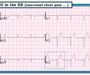
A 50-something with no previous cardiac history and no risk factors presented to the ED with acute chestpain (pressure) that radiated to the left arm. But even without these additional findings — the "Must Recognize" ECG pattern in this patient with new chestpain — is the unmistakeable shape of the ST depression in leads V2 and V3!
Sent by Dan Singer MD, written by Meyers, edits by Smith A man in his late 30s presented with acute chestpain and normal vitals except tachycardia at about 115 bpm. Dr. Singer sent this to me with just the information: "~40 year old with acute chestpain". Anxiety is a common cause of chestpain with tachycardia.
Chris Mondie of the Newark Beth Israel Emergency Medicine Residency sent this case A 50-something man presented with acute chestpain. Among the remarkable chest lead ECG findings include: As per Dr. Mondie — the T waves in leads V 2 and especially in V 3 “tower over” the respective R waves in these leads.
Written by Pendell Meyers, with edits by Steve Smith A man in his early 40s with history of MI s/p PCI presented with bilateral anterior chestpain described as burning and belching with no radiation since last night starting around 11pm (roughly 11 hours ago). The patient was still with ongoing chestpain at the time ECG #1 was done.
Written by Jesse McLaren, with comments from Smith and Grauer A 60 year old presented with three weeks of intermittent non-exertional chestpain without associated symptoms. A prospective validation of the HEART score for chestpain patients at the emergency department. JAMA Intern Med 2019 9. Int J Cardiol 2013 2.
He then went on to say: "40-something with chestpain for one hour. Burning pain subxiphoid and into throat." This is also true in lead III.) == Comment by K EN G RAUER, MD ( 4/22/2019 ): == I liked this case — because it provides unique insight on recognition of a subtle abnormality. Sounds like reflux, right?
== MY Comment by K EN G RAUER, MD ( 9/17/2020 ): == Todays patient is a previously healthy, 60-something year-old woman who presented with chestpain that began at a reception. We are indebted to Dr. Smith for developing Modified Smith-Sgarbossa Criteria for assessing ST-T wave changes in chestpain patients with LBBB.
Written by Pendell Meyers, edits by Smith and Grauer A man in his late 20s with history of asthma presented to the ED with a transient episode of chestpain and shortness of breath after finishing a 4-mile run. His symptoms of chestpain and shortness of breath were attributed to an asthma exacerbation during exercise.
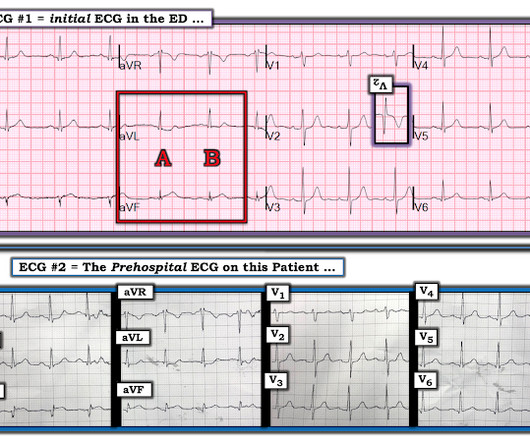
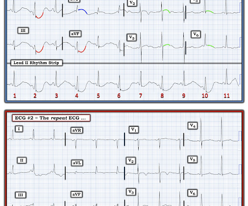
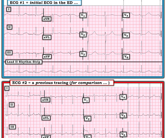
For full discussion of this case — See ECG Blog #292 — == The 2 ECGs shown in Figure-1 were obtained from a man in his 30s — who presented to the ED ( E mergency D epartment ) with chestpain that began several hours earlier. ECG #2 was recorded 1 hour after ECG #1. Initial troponin was negative.
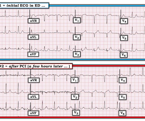
It is from a 50-something with chestpain: What do you think? Up until recently — all computerized ECG interpretation programs that I am aware of used standard millimeter-based STEMI critieria as the basis for determining which chestpain patients should "qualify" for prompt cath with PCI. This was sent to me by a friend.
A 40-something woman with h/o obesity, hyperlipidemia, smoking, as well as cocaine and alcohol abuse presented with intermittent chestpain for 2-3 weeks that became constant in the last day. I illustrate use of a Systematic Approach to ECG Interpretation in My Comment at the bottom of the June 28, 2019 SSmith Post ).
He presented to the ED because he developed sudden severe, sharp, pleuritic (but not positional), substernal and left mid to lower chestpain. Another similar case: Teenager with chestpain and slightly elevated troponin. 13, 2019 Dr. Smith post. 27, 2019 Dr. Smith post. What happens then? Pericarditis?
Methods We prospectively included 1384 patients with LRHSs between March 2019 and March 2021. Therefore, this study aims to investigate the value of NISI in risk stratification and predicting cardiac events in patients with low-risk HEART scores (LRHSs).
As discussed in detail in ECG Blog #228 — this seemingly qualifies as a “ Silent ” MI ( Approximately half of those MIs not accompanied by CP — have some other associated symptom such as syncope, which substitutes as a “chestpain equivalent” ). A picture is worth 1,000 words. What is M INOCA? The November 15, 2023 post in Dr.
The main heart attack symptoms assessed by the study were: Chestpain or discomfort Shortness of breath Pain or discomfort in arms or shoulders Feeling weak, lightheaded, or faint Jaw, neck, or back pain Reference Mahajan S, Valero-Elizondo J, Khera R, Desai NR, Blankstein R, Blaha MJ, Virani SS, Kash BA, Zoghbi WA, Krumholz HM, Nasir K.
For additional examples of this phenomenon whereby automatic truncation of an EMS ECG may mistakenly result in LVH serving to mimi c ischemia — Check out My Comment at the bottom of the page of the following posts in Dr. Smith's ECG Blog — the November 29, 2023 post — June 20, 2020 — March 31, 2019 — March 29, 2019 — and t he December 27, 2018 post.
He woke up alert and with chestpain which he also had experienced intermittently over the previous few days. The history in today's case with sudden loss of consciousness followed by chestpain is very suggestive of ACS and type I ischemia as the cause of the ECG changes. What do you think?
Written by Pendell Meyers A man in his early sixties with no significant medical history (including a "negative cardiac workup a few years ago" for unclear indication) called 911 for acute chestpain constantly for the past 5 hours. However, this patient has active chestpain, and thus this this is inferior-posterior OMI. .:
The ECG in Figure-1 was obtained from a 90-year old woman — who presented with a 2-to-3 day history of chestpain, that became worse on the day of admission. That said — the important point is her ongoing ( and increasing ) chestpain. C ASE F ollow- U p: The patient's chestpain was controlled.
We organize all of the trending information in your field so you don't have to. Join thousands of users and stay up to date on the latest articles your peers are reading.
You know about us, now we want to get to know you!
Let's personalize your content
Let's get even more personalized
We recognize your account from another site in our network, please click 'Send Email' below to continue with verifying your account and setting a password.


Let's personalize your content