This site uses cookies to improve your experience. To help us insure we adhere to various privacy regulations, please select your country/region of residence. If you do not select a country, we will assume you are from the United States. Select your Cookie Settings or view our Privacy Policy and Terms of Use.
Cookie Settings
Cookies and similar technologies are used on this website for proper function of the website, for tracking performance analytics and for marketing purposes. We and some of our third-party providers may use cookie data for various purposes. Please review the cookie settings below and choose your preference.
Used for the proper function of the website
Used for monitoring website traffic and interactions
Cookie Settings
Cookies and similar technologies are used on this website for proper function of the website, for tracking performance analytics and for marketing purposes. We and some of our third-party providers may use cookie data for various purposes. Please review the cookie settings below and choose your preference.
Strictly Necessary: Used for the proper function of the website
Performance/Analytics: Used for monitoring website traffic and interactions
Image courtesy of Takenobu Shimada, Osaka Metropolitan University (CC BY 4.0, [link] mtaschetta-millane Mon, 07/29/2024 - 09:09 July 29, 2024 — When it comes to treating cardiacarrest, acting quickly can mean the difference between life and death. Cardiacarrest can lead to death within minutes.
Out-of-hospital cardiacarrest survival rates dropped significantly at the onset of the COVID-19 pandemic in 2020 and have continued to remain lower than in the pre-pandemic years of 2015–2019, according to a preliminary study to be presented at the American Heart Association's Scientific Sessions 2024.,
Every 10 years, the American Heart Association (AHA) Emergency Cardiovascular Care Committee establishes goals to improve survival from cardiacarrest. Circulation, Ahead of Print.
A patient had a cardiacarrest with ventricular fibrillation and was successfully defibrillated. Coronary Angiography after CardiacArrest without ST-Segment Elevation. N Engl J Med [Internet] 2019;Available from: [link] Should all patients with shockable arrest be taken to angiography regardless of STEMI or No STEMI?
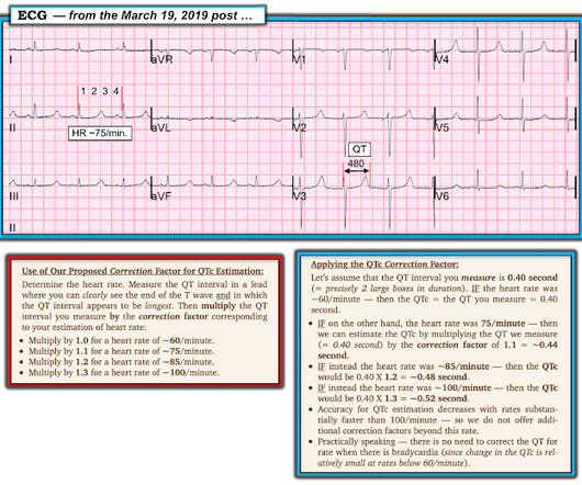
See this post: How a pause can cause cardiacarrest 2. Applying my method to the March 19, 2019 case that I show in Figure-1 the rhythm in this Figure-1 ECG is regular, with an R-R interval just under 4 large boxes. The plan: 1. Place temporary pacemaker 3. Discontinue amiodarone, since it prolongs the QT 4. J Am Coll Cardiol.
Shortly after arrival in the ED ( E mergency D epartment ) — she suffered a cardiacarrest. BUT — Cardiac catheterization done a little later did not reveal any significant stenosis. Figure-1: The initial ECG in today's case — obtained after successful resuscitation from cardiacarrest. ( No CP ( C hest P ain ).
Background Data on the management of patients with cancer presenting with sudden cardiacarrest (SCA) are scarce. Methods Prospective, population-based registry including every out-of-hospital SCA in adults in Paris and its suburbs, between 2011 and 2019, with a specific focus on patients with cancer.
BackgroundTelecommunicator CPR (T‐CPR), whereby emergency dispatch facilitates cardiacarrest recognition and coaches CPR over the telephone, is an important strategy to increase early recognition and bystander CPR in adult out‐of‐hospital cardiacarrest (OHCA). Journal of the American Heart Association, Ahead of Print.
Cardiovascular Implications of Fatal Outcomes of Patients With Coronavirus Disease 2019 (COVID-19). Association of Coronavirus Disease 2019 (COVID-19) With Myocardial Injury and Mortality. Cardiac troponin I in patients with coronavirus disease 2019 (COVID-19): Evidence from a meta-analysis. Guo T, Fan Y, Chen M, et al.
The ECG in Figure-1 — was obtained from a middle-aged man who presented to the ED ( E mergency D epartment ) in cardiacarrest. I i llustrate the ECG finding of T-QRS-D below in Figure-3 , which I've excerpted from My Comment in the November 14, 2019 post in Dr. Smith's ECG Blog. Should you activate the cath lab?
Some patients have baseline RBBB with LAFB, but in patients with likely ACS, these are associated with severe infarction with cardiacarrest, cardiogenic shock or impending shock. In Figure-1 — I reproduce major points that I've summarized from Dr. Smith's August 9, 2019 post on the subject.
The ECG in Figure-1 was obtained from an 18-year old woman — who moments before been resuscitated from out-of-hospital cardiacarrest. Does this ECG in Figure-1 provide clue(s) to the etiology of this patient's cardiacarrest? I suspected the answer resides in the reason why an 18-year woman might have a cardiacarrest.
Two hours later , the patient returned to this same urgent care facility with worsening chest pain, and this ECG was performed: While getting another ECG, the patient suffered cardiacarrest: After multiple defibrillations, ROSC was achieved with ongoing "STEMI". Clinical Cardiology 2019. Long term follow up is unavailable.
We periodically review this intriguing ECG finding that is best known for its association with hypothermia — but which may also be seen in association with a number of other entities, including acute infarction and cardiacarrest. My Comment addresses a few additional aspects of this phenomenon.
Methods Patients who developed LVFWR following AMI and underwent surgical repair at our Institution from January 1990 to December 2019 were considered. Low cardiac output syndrome was the main cause of postoperative death. The aim of this study was to report a single-center experience in this field over a period of 30 years.
Method This is a retrospective cohort study of consecutive ambulance attendances for non-traumatic shock in Victoria, Australia (January 2015–June 2019) linked with government-held administrative data (emergency, admissions and mortality records). Predictors of 30-day mortality were assessed using Cox proportional regressions.
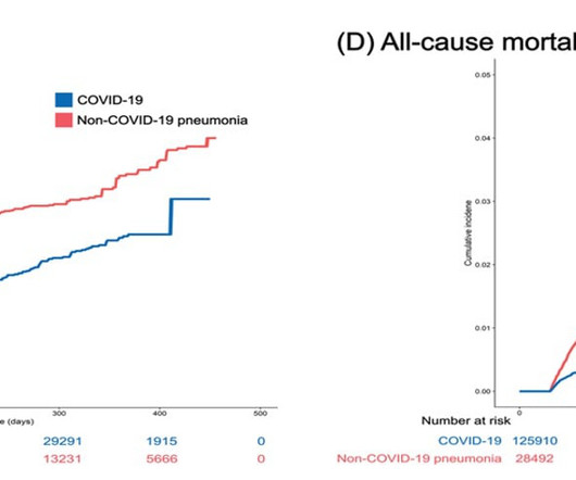
Researchers analyze primary and secondary cardiovascular outcomes in 132,784 inpatients with COVID-19 (October 8, 2020 to September 30, 2021) and 31,173 inpatients with non-COVID-19 pneumonia (January 1, 2019 to December 31, 2019) in Korea. The results indicate a lower risk of cardiovascular disease in COVID-19 patients.
This patient is actively dying from a left main coronary artery OMI and cardiacarrest from VT/VF or PEA is imminent! Complete LMCA occlusion is associated with clinical shock and/or cardiacarrest. The arterial blood gas showed a lactic acidosis with a lactate level of 17mmol/L.
Blood was drawn , and the patient was promptly placed in a room to be seen — but on entering, the ED physician found her unresponsive in cardiacarrest. Do you see any indication on this ECG of WHY this patient was about to arrest? Is there any indication on this ECG of WHY this patient shortly after had a cardiacarrest?
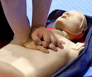
Using automated external defibrillators (AEDs) and cardiopulmonary resuscitation (CPR) as soon as possible increases a person's chance of surviving a cardiacarrest. After meeting the exclusion criteria, more than 9,500 cases of out-of-hospital cardiacarrest were included in the study cohort. versus 4.6%
Polymorphic Ventricular Tachycardia Long QT Syndrome with Continuously Recurrent Polymorphic VT: Management CardiacArrest. A New Seizure in a Healthy 20-something More cases of long QT not measured correctly by computer (these are all fascinating ECGs/cases): Bupropion Overdose Followed by CardiacArrest and, Later, ST Elevation.
Figure-2: A rapid method for estimating the QTc ( Figure from My Comment in the March 19, 2019 post in Dr. Smith's ECG Blog ). == Clinical Implications of a Short QTc: The differential diagnosis for today's tracing, with its short QTc ~360 msec. ) — but as can be seen, my estimate of ~360 msec. mg/dL — vs normal values = 4.4-5.2
And pretty much every doctor can recall an event where a patient experienced a suddenly stressful event and had a cardiac event. That event might have been a heart rhythm issue or even a cardiacarrest. In the hours after the 911 attacks on the World Trade Centre in New York, the rates of cardiacarrest more than doubled.
We provide contemporary nationwide estimates of the temporal trends of SCD in young individuals (135 years of age) from 2000 through 2019 and correlate these trends to changes in out-of-hospital cardiacarrest (OHCA) patterns, rates of inherited cardiac diseases, and implantations of implantable cardioverter defibrillators (ICD).METHODS:All
We analyze the national trends in mechanical thrombectomy use and outcomes for stroke five years after publication of the US guideline update.MethodsWe analyzed the National Inpatient Sample from 2012‐2019. ICD‐9 and ICD‐10 codes identified Ischemic stroke and mechanical thrombectomy patients.
Figure-2: Reasons for the varied ECG presentation of acute LMain occlusion — excerpted from Dr. Smith’s 8/9/2019 post ( See text ). == Addendum Note: Quinidine has a long history of use to treat cardiac arrhythmias and severe malaria. Figure-1: I've labeled the initial ECG in today's case. (
We sought to describe current treatment selection patterns and hospitallevel variability.Methods and ResultsWe identified patients presenting with STEMI with a culprit lesion on coronary angiography between January 1, 2019, and March 31, 2023, using the NCDR (National Cardiovascular Data Registry) CPMI (Chest PainMyocardial Infarction) registry.
This study evaluates the outcomes in patients admitted for HF with and without hyperkalemia.Methods:We used the Nationwide Readmissions Database (NRD) from 2016-2019, extracting adult patients with a primary diagnosis of HF who were admitted between January and November of each year. Survey procedures were applied using SAS 9.4.Results:We
Patients who had ECMO initiated within 24 hours (n=5882 [68.2%]) differed from those who had ECMO initiated after 24 hours, with younger age, more preceding cardiacarrest, and worse acidosis. Multivariable logistic regression evaluated the association between time from admission to ECMO initiation and in‐hospital death.
A secondary analysis evaluated outcomes for severe HF hospitalizations (cardiogenic shock, cardiacarrest, and mechanical ventilation). Centers performing at least 1 heart transplant or left ventricular assist device were classified as ATCs. of centers), 525 037 (18.3%) were admissions to ATCs (5.5%
Osborn waves have been reported with hypercalcemia, brain injury, subarachnoid hemorrhage, Brugada syndrome, cardiacarrest from VFib — and — severe, acute ischemia resulting in acute MI ( See My Comment in the November 22, 2019 post on Dr. Smith’s Blog ). Rituparna et al — as well as Chauhan and Brahma ( Int.
mg experienced a 23% lower incidence of death from cardiovascular causes, resuscitated cardiacarrest, myocardial infarction, stroke, or urgent hospitalization for angina leading to coronary revascularization in a time-to-event analysis. 2019 Sep 10;140(11):e649-e650] [published correction appears in Circulation. References: 1.
Further research and attention to this area are crucial for improving patient outcomes and guiding clinical interventions in this challenging condition.MethodsICD‐10‐CM codes were used to query the National Inpatient Sample (NIS) for patients with AIS between 2010 and 2019. Patients with AKI were on average older (63.29
The 2019 ESC Guidelines for the management of patients with supraventricular tachycardia indicated that IV Amiodarone should not be considered in these populations. Regarding AFib with WPW: The very rapid heart rate and at times extremely short R-R intervals put the patient with AFib and WPW at risk of cardiacarrest from VFib.
Unfortunately — the patient abruptly developed hypoxemia, followed by cardiacarrest with PEA. It was thought that this action precipitated the patient's desaturation, and led to his cardiacarrest. The plan was to proceed as soon as possible with aortic valve replacement. He could not be resuscitated.
But the full cohort of the CABANA trial did not show a significant reduction in the primary composite end point of death, disabling stroke, serious bleeding or cardiacarrest [7]. 2019 Apr 2;321(13):1261-1274. EAST-AFNET 4 trial had 2789 patients with early atrial fibrillation and cardiovascular conditions [8].
Subendocardial Ischemia from another Cause ( ie, sustained tachyarrhythmia; cardiacarrest; shock or profound hypotension; GI bleeding; anemia; "sick patient" , etc. ). The January 9, 2019 post in Dr. Smith's ECG Blog ( Please scroll down to the bottom of the page to see My Comment ).
Am J Med 2019, 132(5):622-630. Now there is a paper published in 2019 that proves the point beyond doubt, though makes it clear that this pattern is associated with very high mortality. American Journal of Medicine 132(5):622-630; May 2019. J Electrocardiol 2013;46:240-8 2. Harhash AA, Huang JJ, Reddy S, et al.
There was 100% proximal LAD occlusion with TIMI 0 flow, and cardiacarrest in the cath lab. Finally — Note that there is also T-QRS-D ( T erminal QRS D istortion ) in lead V3 ( See My Comment in the November 14, 2019 post for review and illustration of T-QRS-D criteria ).
This ECG pattern may be diagnostic of B rugada S yndrome IF seen in association with: i ) a history of cardiacarrest; polymorphic VT; or of non-vagal syncope; and / or ii ) a positive family history of sudden death at an early age; and / or iii ) a similar ECG in relatives. This is not RBBB ( TOP LEFT in Figure-2 ).
No more abnormal U-waves == MY Comment, by K EN G RAUER, MD ( 11/18/2019 ): == LOTS of great points regarding use of the ECG in association with electrolyte abnormalities. From EMCrit: Taking control of severe hyponatremia with DDAVP An ECG recorded 2 days later with a K of 4.1: Note increasing U wave amplitude ( See text ).
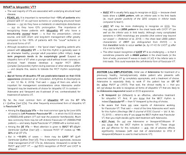
Not all forms of idiopathic VT are predictable based on their ECG appearance ( Anderson et al, 2019 ). Anderson et al, 2019 ) — the chest lead LBBB pattern characteristic of LVOT ( as distinguished from RVOT ) — is that transition tends to occur earlier ( ie, by V1-V2 for LVOT vs after V3 or V4 for RVOT ).
2017 AHA/ACC/HRS guideline for management of patients with ventricular arrhythmias and the prevention of sudden cardiac death. Heart Rhythm, 15(9): 1394-1401. [7] 7] American College of Cardiology/American Heart Association Task Force on Clinical Practice Guidelines and the Heart Rhythm Society.
These include ( among others ) — acute febrile illness — variations in autonomic tone — hypothermia — ischemia-infarction — malignant arrhythmias — cardiacarrest — and especially Hyperkalemia. Patients with such conditions that may transiently mimic the ECG findings of a Brugada-1 pattern are said to have Brugada Phenocopy.
We organize all of the trending information in your field so you don't have to. Join thousands of users and stay up to date on the latest articles your peers are reading.
You know about us, now we want to get to know you!
Let's personalize your content
Let's get even more personalized
We recognize your account from another site in our network, please click 'Send Email' below to continue with verifying your account and setting a password.


Let's personalize your content