This site uses cookies to improve your experience. To help us insure we adhere to various privacy regulations, please select your country/region of residence. If you do not select a country, we will assume you are from the United States. Select your Cookie Settings or view our Privacy Policy and Terms of Use.
Cookie Settings
Cookies and similar technologies are used on this website for proper function of the website, for tracking performance analytics and for marketing purposes. We and some of our third-party providers may use cookie data for various purposes. Please review the cookie settings below and choose your preference.
Used for the proper function of the website
Used for monitoring website traffic and interactions
Cookie Settings
Cookies and similar technologies are used on this website for proper function of the website, for tracking performance analytics and for marketing purposes. We and some of our third-party providers may use cookie data for various purposes. Please review the cookie settings below and choose your preference.
Strictly Necessary: Used for the proper function of the website
Performance/Analytics: Used for monitoring website traffic and interactions
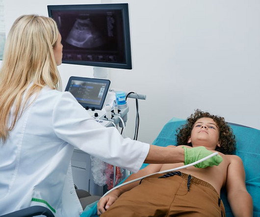
Food and Drug Adminstration (FDA) has approved DEFINITY (Perflutren Lipid Microsphere) as an ultrasound enhancing agent for use in pediatric patients with suboptimal echocardiograms, including those who have undergone heart transplant, or have Kawasaki disease or a congenital cardiovascular anomaly. Lantheus announced that the U.S.
BACKGROUND:Prior clinical trials have demonstrated the efficacy of ultrasound-facilitated catheter-directed thrombolysis (USCDT) for the treatment of acute intermediate-risk pulmonary embolism (PE) using reduced thrombolytic doses and shorter infusion durations. Circulation: Cardiovascular Interventions, Ahead of Print.
The algorithm uses deep learning to analyse routine ultrasound scans of the heart ( echocardiograms ) to detect disease that often goes undetected during standard assessments. For more information: [link] References: 1 Maurer M, Elliott P, Comenzo R, et al. Circulation. 135(14):1357-1377. 2 Siddiqi O., Trends Cardiovasc Med.
Consequently, Philips is well positioned to support CVI treatment by offering a robust portfolio of medical technology that includes both intravascular ultrasound and a differentiated venous stenting system.” 2018 Apr 17;2(2):199-208. Venography versus intravascular ultrasound for diagnosing and treating iliofemoral vein obstruction.
“Trends over time demonstrate the incidence of postoperative VTE to be largely unchanged over the 12-year study period; however, the associated mortality rate slowly decreased from 20% in 2009 to 8% in 2018,” said Dr. Axtell.
Methods We analyzed a cohort of patients admitted for ACS between February 2017 and February 2018. A pulmonary ultrasound was performed on admission and was considered positive (PE+) when there were three or more B-lines in two quadrants or more of each hemithorax. The sensitivity of an NT-proBNP value more than 3647 was 88.9% (51.9–99.7%),
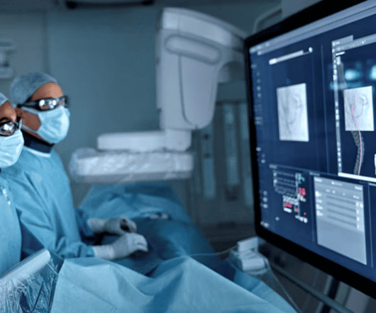
The LumiGuide is actually Philips’ second generation FORS-based imaging system, building upon a first gen FORS system that launched for (very) limited clinical use in 2018. Philips was sure to contrast these capabilities versus X-ray guidance, particularly noting X-ray’s radiation risks, and ability to only produce 2D black and white images.
Intravascular imaging (IVI), such as intravascular ultrasound (IVUS) and optical coherence tomography (OCT), play a crucial role in assessing lesion characteristics and optimizing stent placement during percutaneous coronary intervention (PCI). Patients were divided into two groups based on IVI usage.
Calcified, Non Calcified, Mixed (A combination of the two) Calcified, Fibrous, Non-Calcifed (Fibrofatty + Necrotic Core plaque) Cleveland Clinic Journal Of Clinical Medicine Sept 2018. 4 Coronary atheroma regression and plaque characteristics assessed by grayscale and radiofrequency intravascular ultrasound after aerobic exercise.
A formal ultrasound later showed reasonably good LV function, and so he later received carvedilol and diltiazem, Unfortunately, those led to hypotension at 80/40 with a HR 40. Several hours later, this was the effect: NT pro-BNP elevated to 7000 Furosemide was also given.
Case continued A bedside ultrasound showed diminished LV EF and of course bradycardia. For review — GO TO: The June 4, 2018 post ( LA-LL reversal ). The July 29, 2018 post ( LA-RA reversal ). The November 4, 2018 post ( Leads V1,V2 misplacement ). When narrow (above His bundle), it is likely to be atropine responsive.
A b rief chart review revealed his most recent echo in 2018, with LV EF 67%, “very small” inferior wall motion abnormality. Pads were placed with ultrasound guidance, so they were in the correct position. Over past 3 months, he has had similar intermittent episodes of sharp chest pain while running, but none at rest. What to do now?
This was a point of care ultrasound, not a bubble contrast echo. Nevertheless, this ECG pattern of subendocardial ischemia needs to be instantly recognized — so that optimal decision-making based on the clinical scenario can promptly begin ( See the October 31, 2018 post on Dr. Smith’s Blog ). First trop I returns at 1.5.
My Comment, by KEN GRAUER, MD ( 6/17/2018 ): = Excellent case with insightful learning points explaining why these serial tracings are not indicative of acute inferior infarction. Conclusion: 0 0 1 405 2312 MMRF 19 5 2712 14.0 I’ll add the following 2 comments: i ) This patient presumably has effusive-constrictive pericarditis.
A bedside ultrasound should be done to assess volume and other etiologies of tachycardia, but if no cause of type 2 MI is found, the cath lab should be activated NOW. The January 30, 2018 post — for PTA. Smith comment: this is diagnostic of OMI until proven otherwise. The September 22, 2019 post — intermittent ST-T wave artifact.
Journal of Electrocardiology 2018. However, in almost every case, one should confirm absence of OMI (Occlusion MI) at least by contrast ultrasound. Journal of Emergency Medicine 2006; 31(1):67-77. 40-50% of LAD occlusion have zero reciprocal ST depression. 0 0 1 24 140 MMRF 1 1 163 14.0 That may be the case. Smith & Meyers.
The ways to tell for certain include intravascular ultrasound (to look for extra-luminal plaque with rupture) or "optical coherence tomography," something I am entirely unfamiliar with. The authors recommend using optical coherence tomography or intravascular ultrasound imaging in patients with evidence of nonobstructive CAD by angiogram.
Here was his prehospital ECG, which I viewed immediately while the resident performed cardiac ultrasound: What do you think? Here is the cardiac ultrasound which the resident performed as I viewed the ECG: This shows a huge pericardial effusion. Therefore, we performed ultrasound-guided pericardiocentesis. Is is sinus?
You use an ultrasound. Metabolic Syndrome and Related Disorders , 2018; 6 Application of non-HDL cholesterol for population-based cardiovascular risk stratification: results from the Multinational Cardiovascular Risk Consortium. Regardless of the murmur findings they describe. Which can now be used easily at the bedside.
Many of these issues were described in a prior post by Dr. Angie Lobo ( @aloboMD ) (For open-access reviews of this literature, see Saw 2016 , Saw 2017 , or Hayes 2018.) Often, intravascular ultrasound or intravascular optical coherence tomography is requeried to make the diagnosis.
Can J of Cardiol 2018, 34: 132-145 Here are some other cases: LVH, LBBB, RBBB, and RVH may manifest ST depression without any ischemia! Methods STEMI activations between January 2014 and April 2018 at the University of Arizona Medical Center were identified. A emergent cardiology consult can be helpful for equivocal cases.
This was diagnosed by IVUS (intravascular ultrasound) as a ruptured plaque. My Comment , by K EN G RAUER, MD ( 10/24/2018 ): = Important teaching points are made in this post by Dr. Smith. Values: STE60V3 = 2.0, QRS V2 = 10, RAV4 = 15.5, QTc = 377 by computer 4-variable formula value = 16.2, There was good flow. It was stented.
My bedside ultrasound was of insufficient quality, but showed somewhat reduced overall EF, distended IVC without respiratory variation, no pericardial effusion, and diffuse bilateral B lines. == What do you think of her ECG? J Electrocardiol, 42 (2009), pp.
In 2018, he even opened a state of the art lab nicknamed “the Watcher” and was paid $18 million from Medicare in the next 3 years. A lower extremity arterial ultrasound revealed elevated velocities in the right proximal superficial femoral artery. 12.27.2016. Mr. Rosenberg meets Dr. Dormu.
Her bedside cardiac ultrasound was normal We decided to cardiovert her since the time of onset was very recent. For more on SSS — See My Comment at the bottom of the page in the July 5, 2018 post in Dr. Smith’s ECG Blog. But when you see this, you should suspect that the AV node is not well. I signed her out to one of my partners.
His ED cardiac ultrasound (which is not at all ideal for detecting wall motion abnormalities, and is also very operator dependent for this finding) was significant for depressed global EF. The patient's initial troponin I was 2.0 ng/mL (99% reference level = 0.030 ng/mL. With his ESRD, he does have an elevated baseline troponin at ~0.40
ALL TROPS WERE UNDETECTABLE A formal ultrasound was done: Normal estimated left ventricular ejection fraction at rest. For another case in which marked ST elevation in leads V1 and V2 could easily be mistaken for a hyperacute change — See the Figure I drew in My Comment at the bottom of the December 27, 2018 post on Dr. Smith’s ECG Blog.
Am J Cardiol 2018; 122(8):1303-1309. This was missed by the physicians, even with a bedside speckle tracking ultrasound: no wall motion abnormality was seen. In summary: At a cutpoint of 17.0, it is 97% sensitive. At a cutpoint of 19.0, it is 97% specific. Finally, there is a simplified formula without QT correction : Aslanger.
A bedside cardiac ultrasound revealed grossly normal to hyperdynamic systolic function with no obvious areas of wall motion abnormalities. Heart Rhythm 2018. This was recorded about 30 minutes later: Same A previous ECG was obtained and was normal. His lab workup was significant for positive influenza A rapid test and hyponatremia.
We organize all of the trending information in your field so you don't have to. Join thousands of users and stay up to date on the latest articles your peers are reading.
You know about us, now we want to get to know you!
Let's personalize your content
Let's get even more personalized
We recognize your account from another site in our network, please click 'Send Email' below to continue with verifying your account and setting a password.


Let's personalize your content