This site uses cookies to improve your experience. To help us insure we adhere to various privacy regulations, please select your country/region of residence. If you do not select a country, we will assume you are from the United States. Select your Cookie Settings or view our Privacy Policy and Terms of Use.
Cookie Settings
Cookies and similar technologies are used on this website for proper function of the website, for tracking performance analytics and for marketing purposes. We and some of our third-party providers may use cookie data for various purposes. Please review the cookie settings below and choose your preference.
Used for the proper function of the website
Used for monitoring website traffic and interactions
Cookie Settings
Cookies and similar technologies are used on this website for proper function of the website, for tracking performance analytics and for marketing purposes. We and some of our third-party providers may use cookie data for various purposes. Please review the cookie settings below and choose your preference.
Strictly Necessary: Used for the proper function of the website
Performance/Analytics: Used for monitoring website traffic and interactions
You cannot eliminate the plaque entirely, but multiple clinical trials have shown plaque regression using high-intensity cholesterol-lowering treatments, which I have discussed previously. All of these parameters are important and need to be considered when evaluating plaque regression. REVERSAL Investigators.
I am excited about the potential of the FAI-Score biomarker, which has promising prognostic value beyond existing CT-based methods such as plaque, calcium scoring, and CAD-RADS based interpretation." fold higher risk for cardiac mortality and 5.5-fold The CaRi-Heart technology is in clinical use in the UK, European Union, and Australia.
FFRCT, coronary plaque, etc). Even with this broader definition, cardiovascular AI’s total share of AI clearances is declining, falling from roughly 25% of clearances in 2018-2019, to 16.5% Cardiovascular AI actually makes up a larger 17.4% in 2020-2022, and 13.5% since the start of 2023.
We've experienced a year of notable accomplishments and growth including the expansion of our product portfolio with Plaque Analysis and RoadMap Analysis. Neth Heart J 2018. HeartFlow is committed to serving customers quickly and reliably with a median turnaround time less than 1.5 For more information: www.heartflow.com References 1.
Introduction:Alzheimers Disease (AD), characterized by extracellular deposition of amyloid beta (A) plaques in brain tissue, is often comorbid with cerebral amyloid angiopathy, which carries an elevated risk of intracranial hemorrhage. Stroke, Volume 56, Issue Suppl_1 , Page ATP205-ATP205, February 1, 2025. andICD-10-CMcode G30.x.
If the arrest was caused by acute MI due to plaque rupture, then the diagnosis is MINOCA. Here is my comment on MINOCA: "Non-obstructive coronary disease" does not necessarily imply "no plaque rupture with thrombus." They often cannot even be recognized as culprits, as fissured or ulcerated plaque. FFR can be useful.
Today, they viewed the angiogram and concluded that the thrombus at the mid RCA must have extended proximally from the culprit ruptured plaque, extending proximal to the RV marginal branch and temporarily occluding it. 24, 2018 ECG Blog — Please scroll down to the bottom of the page to My Comment, in the section "Beyond-the-Core". ).
Here’s the angiogram of the RCA : No thrombus or plaque rupture in the RCA (or any coronary artery) was found. This MI wasn’t caused by a ruptured plaque of CAD - it was a coronary artery dissection of the RCA. Angiography Angiography was performed after aspirin and heparin were started.
Can J of Cardiol 2018, 34: 132-145 Here are some other cases: LVH, LBBB, RBBB, and RVH may manifest ST depression without any ischemia! Methods STEMI activations between January 2014 and April 2018 at the University of Arizona Medical Center were identified. Miranda DF, Lobo AS, Walsh B, et al.
The coronary angiogram revealed no critical stenosis, or acute plaque ulceration. Surawicz and Knilans report that intense catecholamine surge, or severe maladjustment of the autonomic nervous system, can manifest “cerebral T waves” in the absence of an acute intracranial process. Furthermore, pertinent electrolyte values (e.g. Friedman, M.,
It was long ago that I realized that Wellens' is a reperfusion pattern and the pattern A is early and evolves into Pattern B. -- Comment by K EN G RAUER, MD ( 7/31/2018 ): -- In my experience, the most instructive ECG cases consist of serial tracings with contemporaneous notation at each moment in time as the case evolves. 2012;5:134–137.
A cardiac CT is a low-dose CT scan of your heart that assesses whether or not you have plaque in your coronary arteries and, if so, how much. In general, the more plaque you have, the higher your risk of a heart attack over the next 10 years. 2018 Sep 4;72(10):1141-1156. J Am Coll Cardiol. 6 A Test in Context: Lipoprotein(a).
The CAC scan looks for deposits of calcium in the areas of the coronary arteries as a proxy marker for plaque. It tells you ‘ if ’ there is plaque and how much, as a score called a CAC score. About 1 in 10 patients with a CAC of 0 will have some plaque identified on more in-depth scanning using CTCA 1. Int J Cardiol.
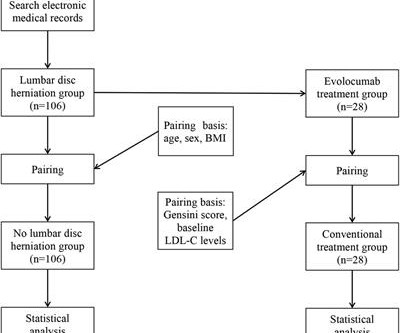
Objective Assessing the impact of lumbar disc herniation (LDH) on the plaque burden of coronary atherosclerosis is our objective. Methods In this study, a total of 212 patients (age 46–80 years) with unstable angina (UA) who underwent coronary angiography (CAG) in our hospital from January 2018 to July 2022 due to UA were included.
Recent evidence suggests that nonstenotic carotid plaque (nsCP) may be a substantial contributor to the risk for ESUS. Stroke, Ahead of Print. BACKGROUND:Many ischemic strokes are diagnosed as embolic strokes of undetermined source (ESUS).
Detailed analysis of the excised carotid plaques were carried out with pyrolysis-gas chromatography-mass spectrometry, stable isotope analysis, and electron microscopy. Primary endpoint of the study was a composite of myocardial infarction, stroke, or death from any cause in those who had micro and nanoplastics in the carotid plaque.
It provides anatomic data, plaque identification and characterization, as well as the calculations of FFR CT , a coronary physiological simulation, computed from simulated pressure, velocity and blood flow information obtained from a 3D computer model generated from static coronary CT images. 301–302, [link]. 3 Secemsky, Eric A.,
BACKGROUND:Sex-specific differences in plaque composition and instability underscore the need to explore circulating markers for better prediction of high-risk plaques. Plaque stability was determined by gold-standard histological classifications. Adipokine, lipid, and immune profiling was conducted.
A cardiac CT is a low dose CT scan of your heart that assesses whether or not you have plaque in your coronary arteries and if so, how much. In general, the more plaque you have, the higher your risk of a heart attack over the next 10 years.
5 High intensity interval training induces beneficial effects on coronary atheromatous plaques – a randomized trial, European Journal of Preventive Cardiology , 2022;, zwac309, 6 FOURIER Steering Committee and Investigators. 2018 Mar;68(668):151-152. 2006 Apr 5;295(13):1556-65. N Engl J Med. 2017 May 4;376(18):1713-1722.
A CTCA provides much more anatomical detail and can identify advanced plaque often missed by CT Coronary Artery Calcium Score scans alone. There are 3 types of coronary atherosclerosis visible on CTCA: Calcified Plaque - Easily Identified on both CT CAC & CTCA scans. Subscribe now How Often Does A CT CAC Scan Miss Plaque?
Since 2018, exceptional validation results showing CaRi-Heart technology’s ability to aid the prediction of heart attacks have been published in leading medical journals including the Lancet , JACC , European Heart Journal , and Cardiovascular Research.
There are multiple possible clinical situations that could account for diffuse subendocardial ischemia that is not due to ACS and plaque rupture. The history in today's case with sudden loss of consciousness followed by chest pain is very suggestive of ACS and type I ischemia as the cause of the ECG changes.
Atherosclerotic cardiovascular disease (ASCVD), caused by plaque buildup in arterial walls, is one of the leading causes of disability and death worldwide.1,2 7 Research has shown inflammation plays a significant role in the development of atherosclerosis and ASCVD,8-10 and even the formation of plaque.11 Published 2018 Feb 6.
Arteries generally narrow and occlude for one of two reasons: The progressive accumulation of plaque. A plaque ruptures, and a clot forms in the artery, thereby occluding it. There are the ‘garden variety’ heart attacks whereby a plaque ruptures in the coronary artery, called a spontaneous heart attack.
This was diagnosed by IVUS (intravascular ultrasound) as a ruptured plaque. As there was ruptured plaque, this is NOT Prinzmetal's angina. It is just as dangerous, as there is a ruptured plaque with thrombus (which lysed) in the proximal LAD. Values: STE60V3 = 2.0, QRS V2 = 10, RAV4 = 15.5, There was good flow. It was stented.
The cause of angina usually involves inadequate blood flow reaching the heart muscle because of significant narrowing of the artery due to plaque buildup. 2018 Jan 6;391(10115):31-40. There are many ways it can present but this is the most common. But coronary stenting is not the only way to reduce symptoms of angina. N Engl J Med.
As an aside, the LCx OMI is a type 2 event, since it is due to supply-demand mismatch from thrombus, and not due to atherosclerotic plaque rupture or erosion). The January 30, 2018 post — for PTA. Then, part of the thrombus embolized into the LCx causing an inferoposterolateral OMI. (As The August 26, 2019 post — baseline artifact.
The axiom of "type 1 (ACS, plaque rupture) STEMIs are not tachycardic unless they are in cardiogenic shock" is not applicable outside of sinus rhythm. Atrial Flutter Mimicking ST Depression Inferolateral ST elevation, vomiting, and elevated troponin My Comment by K EN G RAUER, MD ( 11/26/2018 ): Excellent discussion by Drs.
While the total body seems to do little in determining cholesterol levels, what is more scientifically shocking is slope of the curve between blood LDL levels and plaque burden is rarely linear. 2018 Apr 23;2018:8598054. doi: 10.1155/2018/8598054. LDL is obviously a target against atherosclerosis. Mind you LDL constitutes.000025%
In my review of the literature, there are many articles which purport to demonstrate an acutely increased risk of plaque rupture from emotional stress, but I could not find any credible case reports that were not at least as likely to be takotsubo. Mechanisms of plaque formation and rupture. Coronary plaque disruption.
There were no plaques or stenoses. See the September 14, 2018 post for a nice overview of this subject by Dr. Meyers. A workup was undertaken in search of a cause of the patient's ventricular arrhythmia. As noted above echocardiography was completely normal. CT coronary angiogram showed a hypoplastic RCA and dominant LCx.
We organize all of the trending information in your field so you don't have to. Join thousands of users and stay up to date on the latest articles your peers are reading.
You know about us, now we want to get to know you!
Let's personalize your content
Let's get even more personalized
We recognize your account from another site in our network, please click 'Send Email' below to continue with verifying your account and setting a password.


Let's personalize your content