This site uses cookies to improve your experience. To help us insure we adhere to various privacy regulations, please select your country/region of residence. If you do not select a country, we will assume you are from the United States. Select your Cookie Settings or view our Privacy Policy and Terms of Use.
Cookie Settings
Cookies and similar technologies are used on this website for proper function of the website, for tracking performance analytics and for marketing purposes. We and some of our third-party providers may use cookie data for various purposes. Please review the cookie settings below and choose your preference.
Used for the proper function of the website
Used for monitoring website traffic and interactions
Cookie Settings
Cookies and similar technologies are used on this website for proper function of the website, for tracking performance analytics and for marketing purposes. We and some of our third-party providers may use cookie data for various purposes. Please review the cookie settings below and choose your preference.
Strictly Necessary: Used for the proper function of the website
Performance/Analytics: Used for monitoring website traffic and interactions
Chest Pain Severity Rating Is a Poor Predictive Tool in the Diagnosis of ST-Segment Elevation Myocardial Infarction [link] Abstract Current ST-segment elevation myocardial infarction (STEMI) guidelines require persistent electrocardiogram ST-segment elevation, cardiac enzyme changes, and symptoms of myocardial ischemia.
Electromechanical association: a subtle electrocardiogram artifact. Incredibly , this case was just published in Circulation on January 22, 2018 (thanks to Brooks Walsh for finding this!) 2018; 137: 402-404. 2018; 137: 402-404. Journal of Electrocardiology. 2012;45(1):15-17. doi:10.1016/j.jelectrocard.2010.12.162.
Weve also published the largest study on this question: Emergency Department Code STEMI patients with initial electrocardiogram labeled normal by computer interpretation: a 7-year retrospective review. Emergent cardiac outcomes in patients with normal electrocardiograms in the emergency department. Am J Emerg Med. Am J Emerg Med.
Therefore, we aimed to evaluate an artificial intelligence (AI)-enabled ECG algorithm to predict AF detected by PCT after index stroke.Methods:This retrospective study included all adult patients with ischemic stroke evaluated at Mayo Clinic with baseline electrocardiogram (ECG) and PCT between 2018-2020.
Electromechanical association: a subtle electrocardiogram artifact. This case was published in Circulation on January 22, 2018 (thanks to Brooks Walsh for finding this!) 2018; 137: 402-404. Originally published January 22, 2018 Here is a case from Circulation year 2000 that was misdiagnosed as due to pancreatitis.
Apple has made available in Australia its electrocardiogram app for Series 4, 5 and 6 of its Apple Watch. MARKET SNAPSHOT Apple was the first to launch an ECG app in 2018. An irregular rhythm notification feature that checks atrial fibrillation was also included in the Apple Watch Series 3 and later versions.
A 12-lead electrocardiogram, lead V4R , and leads V7-9 were recorded on admission. For review — GO TO: The June 4, 2018 post ( LA-LL reversal ). The July 29, 2018 post ( LA-RA reversal ). The November 4, 2018 post ( Leads V1,V2 misplacement ). The February 11, 2020 post ( LA-RA reversal ).
Meyers, Weingart and Smith published their OMI Manifesto — in which they extensively document the critically important concept that management of acute MI by separation into a “STEMI” vs “non-STEMI” classification is an irreversibly flawed approach.
International evaluation of an artificial-intelligence- powered electrocardiogram model detecting acute coronary occlusion myocardial infarction. Immediate and early percutaneous coronary intervention in very high-risk and high-risk non-ST segment elevation myocardial infarction patients. Clin Cardiol 2022 4. Herman, Meyers, Smith et al.
Electromechanical association: a subtle electrocardiogram artifact. Incredibly , this case was just published in Circulation on January 22, 2018 (thanks to Brooks Walsh for finding this!) 2018; 137: 402-404. 2018; 137: 402-404. Journal of Electrocardiology. 2012;45(1):15-17. doi:10.1016/j.jelectrocard.2010.12.162.
New insights into the use of the 12-lead electrocardiogram for diagnosing acute myocardial infarction in the emergency department. Can J of Cardiol 2018, 34: 132-145 Here are some other cases: LVH, LBBB, RBBB, and RVH may manifest ST depression without any ischemia! Incidence of an acute coronary occlusion.
New insights into the use of the 12-lead electrocardiogram for diagnosing acute myocardial infarction in the emergency department. Paroxysmal atrioventricular block: Eletrophysiological mechanism of phase 4 conduction block in the His-Purkinje system: A comparison with phase 3 block. Pacing Clin Electrophysiol. 40; 1234-1241.
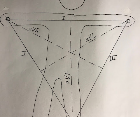
J Electrocardiology January–February, 2018; Volume 51, Issue 1, Pages e5–e6. We have studied the value of the QRS-T angle in normal and abnormal electrocardiograms, and have noted the normal A^QRS ranges between -15^ and +85^ on the hexaxial reference system and locates centrally from normal QRS vectors that vary from -30^ to +95 ^.
DIagnostic accuracy oF electrocardiogram for acute coronary OCClusion resulTing in myocardial infarction (DIFOCCULT study). Eur Heart J 2018 4. Aslanger et al. Int J Cardiol Heart Vasc 2020 3. Lemkes et al. Timing of revascularization in patients with transient ST segment elevation myocardial infarction: a randomized clinical trial.
Below is the post -PCI electrocardiogram. For more on this mirror-image opposite ST-T wave relation in leads III vs aVL — See My Comment in the March 8, 2019 and August 9, 2018 posts in Dr. Smith's ECG Blog ). In the cath lab the patient was found to have a 100% occlusion of a small 1st marginal branch of the LCx.
You will note that it is essentially an unremarkable electrocardiogram except for some PACS. We have often referred to this almost magical m irror- i mage r elationship for ST-T waves in leads I II and aV L — which when present, means a cute i nferior O MI until you prove otherwise ( See My Comment on the 8/9/2018 SSmith Blog Post ).
Meyers, Weingart and Smith in their 2018 OMI Manifesto. Grauer K, Kravitz L, Curry RW, Ariet M: Computerized Electrocardiogram Interpretations: Are They Useful for the Family Physician? NOTE: There is a reason the PM Cardio AI Bot app. has performed so well clinically: It has been programmed using ECG criteria put forth by Drs.
A Deep Neural Network learning algorithm outperforms a conventional algorithm for emergency department electrocardiogram interpretation. This ECG comes from Pierre Taboulet ( [link] /)( [link] ) an ECG whiz who codes a lot of ECGs for Cardiologs' Artificial Intelligence Deep Neural Network algorithm ( [link] ). What an honor.
The stress electrocardiogram is non-diagnostic. For another case in which marked ST elevation in leads V1 and V2 could easily be mistaken for a hyperacute change — See the Figure I drew in My Comment at the bottom of the December 27, 2018 post on Dr. Smith’s ECG Blog. No wall motion abnormality at rest.
Usefulness of the Electrocardiogram in Establishing the Diagnosis and Prognosis of Arrhythmogenic Right Ventricular Cardiomyopathy Other References, from the above article: 1 FI Marcus Epsilon waves aid in the prognosis and risk stratification of patients with ARVC/D J Cardiovasc Electrophysiol, 26 (2015), pp. J Electrocardiol, 42 (2009), pp.
The utility of the triage electrocardiogram for the detection of ST-segment elevation myocardial infarction. October 2018. It is easy to be led astray by the computer. -- My Comment by K EN G RAUER, MD ( 10/9/2018 ): -- I agree with Dr. Smith — This is a faulty study for many reasons. This paper was just published: Noll S.
10 In 2018, the European Society of Cardiology guidelines 11 and the fourth universal definition of myocardial infarction 12 suggested utilizing the original Sgarbossa criteria (Figures 1A to 1C) for diagnosis of occlusion myocardial infarction in both left bundle branch block 13 , 14 and ventricular paced rhythm.15,
New insights into the use of the 12 Lead Electrocardiogram for diagnosing Acute Myocardial Infarction in the emergency department. Readers interested in a more robust discussion of STD vectors, and their implications in OMI, are encouraged to read this phenomenal post at the Smith ECG Blog. link] [1] Mirand, D. 2] Aslanger, E.,
Early Continuous ST Segment Monitoring in Unstable Angina: Prognostic Value Additional to the Clinical Characteristics and the Admission Electrocardiogram. American Journal of Cardiology 2018. (Patel et al., Krucoff et al.) Patel et al. Heart 1996. Krucoff et al. Blondheim et al.
Fever not only unmasks a Brugada-type electrocardiogram (ECG) but also increases the risk of ventricular tachyarrhythmias such as ventricular fibrillation (VF) or sudden cardiac death. Heart Rhythm 2018. Studies have shown a higher prevalence of BrS in febrile patients compared to nonfebrile ones. y (3 of 88, 43.6 ± 37.4
The role of an electrocardiogram (ECG) in routine testing remains controversial in current guidelines. ObjectivesThe occurrence of sudden cardiac death (SCD) in competitive athletes has led to a discussion about appropriate preparticipation screening models. A staged preparticipation screening was performed.
We organize all of the trending information in your field so you don't have to. Join thousands of users and stay up to date on the latest articles your peers are reading.
You know about us, now we want to get to know you!
Let's personalize your content
Let's get even more personalized
We recognize your account from another site in our network, please click 'Send Email' below to continue with verifying your account and setting a password.


Let's personalize your content