This site uses cookies to improve your experience. To help us insure we adhere to various privacy regulations, please select your country/region of residence. If you do not select a country, we will assume you are from the United States. Select your Cookie Settings or view our Privacy Policy and Terms of Use.
Cookie Settings
Cookies and similar technologies are used on this website for proper function of the website, for tracking performance analytics and for marketing purposes. We and some of our third-party providers may use cookie data for various purposes. Please review the cookie settings below and choose your preference.
Used for the proper function of the website
Used for monitoring website traffic and interactions
Cookie Settings
Cookies and similar technologies are used on this website for proper function of the website, for tracking performance analytics and for marketing purposes. We and some of our third-party providers may use cookie data for various purposes. Please review the cookie settings below and choose your preference.
Strictly Necessary: Used for the proper function of the website
Performance/Analytics: Used for monitoring website traffic and interactions
What do you think the echocardiogram shows? Nevertheless, this ECG pattern of subendocardial ischemia needs to be instantly recognized — so that optimal decision-making based on the clinical scenario can promptly begin ( See the October 31, 2018 post on Dr. Smith’s Blog ). NTG drip started. Pain better still.
The algorithm uses deep learning to analyse routine ultrasound scans of the heart ( echocardiograms ) to detect disease that often goes undetected during standard assessments. For more information: [link] References: 1 Maurer M, Elliott P, Comenzo R, et al. Circulation. 135(14):1357-1377. 2 Siddiqi O., Trends Cardiovasc Med.
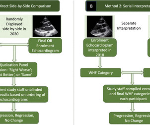
Methods A four-member expert panel reviewed 799 enrolment (in 2018) and completion (in 2020) echocardiograms from the GOAL Trial of latent RHD in Uganda to make consensus determination of normal, borderline RHD or definite RHD. Results There were 799 pairs of echocardiogram assessments included.
Food and Drug Adminstration (FDA) has approved DEFINITY (Perflutren Lipid Microsphere) as an ultrasound enhancing agent for use in pediatric patients with suboptimal echocardiograms, including those who have undergone heart transplant, or have Kawasaki disease or a congenital cardiovascular anomaly. Lantheus announced that the U.S.
That said — distinction between "classic" HCM vs the apical HCM for m may be useful because: i ) ECG findings tend to be different ( Lyon et al — Europace 20:102-112iii, 2018 ) ; — ii ) Echo appearance is different when hypertrophy localizes to the apex; and , iii ) There is a significantly greater incidence of AFib with apical HCM.
Methods Retrospective chart review of 200 patients admitted for ADHF from 2018 to 2019 with transthoracic echocardiogram during index hospitalisation. The aim of this study was to assess the relationship between IVC diameter, clinical variables and ADHF rehospitalisations. Charts were assessed for ADHF rehospitalisation within 1 year.
Two groups of patients' age, gender, diabetes duration, merge disease, echocardiogram and blood biochemical indexes, had no statistical difference (P>0.05). There were no significant differences in the number of coronary artery lesions, treatment regimens, cardiovascular and hypoglycemic drugs between the two groups (P>0.05).
An artificial intelligence enhanced ECG (AIECG) algorithm can predict LVDD and mortality in general populations but has not been examined in cardiac intensive care units.MethodsThis historical cohort study included consecutive adults admitted to Mayo Clinic cardiac intensive care unit from 2007 to 2018 with an admission AIECG.
Results 48 patients (71% with a Fontan circulation, 42% females, mean age 33±9 years) underwent two CPETs between May 2018 and May 2022 with echocardiograms performed within 6 months of each CPET. Apple Watch was the predominant smartwatch used (79%).
5 years ago Similar Previous formal echocardiogram Inferior posterior with dyskinesis "Dyskinesis" is the technical echo term for LV aneurysm. 2 prior ECGs were found in his medical record — the latest of which was done circa 2018 ( which would be ~8 years after his inferior MI — and ~5 years before ECG #1 ).
Another important role is for detection of coronary anomalies, which can also be seen on echocardiogram sometimes. 2018 Jun 28;3:e72-e79. Finding out the origin and insertion of MAPCAs is important prior to surgical repair of tetralogy of Fallot. Magnetic resonance imaging is another way of documenting coronary anomalies. 2018.76824.
Hopefully a repeat echocardiogram will be performed outpatient. Systolic function normal by visual assessment only, unable to visualize well for further characterization. 1900: RBBB and LAFB are almost fully resolved. 2300: QRS now within normal limits. No other significant injuries were found. The patient did well and was discharged.
Eur Heart J 2018. First troponin I returns at 48 ng/L ECG 5 143 min No significant change ECG 6 261 min Same hs Troponin I profile (peaked at 1849): Formal Echocardiogram SUMMARY The estimated left ventricular ejection fraction is 74 %. Full text link. The estimated pulmonary artery systolic pressure is 27 mmHg + RA pressure.
Due to limitations of echocardiogram in evaluating the right ventricle, magnetic resonance imaging study of the right ventricle along with that of the left ventricle has been reported. 2018 Sep;27(9):1052-1062. 2018 Sep;27(9):1063-1071. Effect of exercise on right ventricle. N Engl J Med. 1991 Jan 31;324(5):295-301. Circulation.
The patient underwent an emergent formal echocardiogram to look for wall motion abnormality: The estimated left ventricular ejection fraction is 63 %. Exclusion criteria were age less than 18, SBP less than 100 mmHg, echocardiogram with EF less than 50%, STEMI, pregnancy, and trauma. No wall motion abnormality.
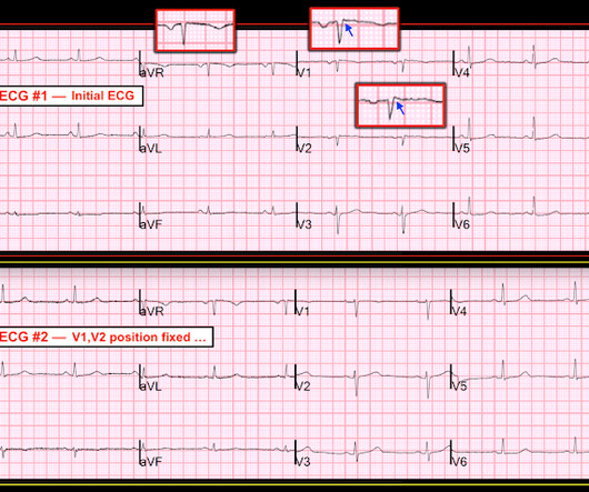
American Journal of Emergency Medicine 36(5):865-870; May 2018. She had a normal echocardiogram, with normal shortening and thickening of the septum. My Comment, by K EN G RAUER, MD ( 11/4/2018 ): = Important post by Dr. Smith regarding recognition of chest lead misplacement of leads V1 and V2. 27, 2018 blog post ).
Formal echocardiogram showed normal EF, no wall motion abnormalities, no pericardial effusion. 72; Issue 9; 2018 ) — A ) Brugada-1 ECG pattern, showing coved ST-segment elevation ≥2 mm in ≥1 right precordial lead, followed by a negative T-wave. — No more troponins were done. He was found to be influenza positive.
Troponins, echocardiogram An echocardiogram showed inferobasilar hypokinesis, further supporting a diagnosis of regional ischemia , likely of the area supplied by the RCA. The biphasic T wave is consistent with recent reperfusion of an occluded coronary artery supplying the inferior region. The initial troponin I was elevated at 0.75
While awaiting transfer to the cath lab, STAT echocardiogram was performed and showed LVEF 30-35%, as well as anterior, inferior, and apical hypokinesis, and apical thrombus. The January 30, 2018 post — for PTA. This confirms the suspicion of prior anterior OMI. The thrombus is circled below in red.
See this case: what do you think the echocardiogram shows in this case? Can J of Cardiol 2018, 34: 132-145 Here are some other cases: LVH, LBBB, RBBB, and RVH may manifest ST depression without any ischemia! Methods STEMI activations between January 2014 and April 2018 at the University of Arizona Medical Center were identified.
An echocardiogram was done. 72; Issue 9; 2018 ) — A ) Brugada-1 ECG pattern, showing coved ST-segment elevation ≥2 mm in ≥1 right precordial lead, followed by a negative T-wave. — Is there also Brugada? Here is the result: The estimated left ventricular ejection fraction is 50 %. Right ventricular prominence.
Indeed, bedside Echocardiogram revealed severe left ventricular impairment of Takotsubo cardiomyopathy. Surawicz and Knilans report that intense catecholamine surge, or severe maladjustment of the autonomic nervous system, can manifest “cerebral T waves” in the absence of an acute intracranial process. potassium) were within normal parameter.
Next day echocardiogram showed inferolateral hypokinesia with an EF of %45-50. On echocardiogram you will not see a "posterior" hypokinesia (will see "inferolateral") and, as in this case, LCx may not give the blood supply of basal inferior segment (formerly called "posterior"). The patient recovered well.
Unfortunately there is no echocardiogram accessible because the patient checked himself out of the hospital in order to get back to his home state before it could be completed. Slow TIMI 2 initially with brisk flow status post percutaneous coronary intervention with 18mm drug-eluting stent. To our knowledge, the patient did well.
I have ordered an echocardiogram which will be done today, after that patient can be discharged to home with follow-up in 2 to 3 months." Meyers, Smith; Weingart wrote an extensive review on Idiopathic VT in the September 14, 2018 post of Dr. Smith’s ECG Blog. 14, 2018 post adds a series of PEARLS on “My Take” regarding this subject.
These are reperfusion T-waves (the same thing as Wellens' waves) Echocardiogram Regional wall motion abnormality-distal septum and apex. Regional wall motion abnormality-distal inferior wall. ECG recorded at time 38 hours: A further evolutionary stage of T-wave inversion. Circ Cardiovasc Interv. 2012;5:134–137. 111.966630. Hayes, S.N.,
A formal echocardiogram was completed the next day and again showed a normal ejection fraction without any focal wall motion abnormalities to suggest CAD. Cardiology was consulted and they agreed that the EKG had an atypical morphology for STEMI and did not activate the cath lab. Heart Rhythm, 4(2), 198-199. [6]
No further echocardiograms were available after cath. The patient was discharged one day after intervention and appears to be doing well. The full thickness infarction with LV aneurysm morphology places him at a higher risk for short and long term complications (e.g., I still at times forget to look for it.
I think a good start would be a posterior EKG and a high quality contrast echocardiogram read by an expert. His prior EF from an ECHO 6 months prior indicated 35% LVEF. What would you do in this scenario? Unfortunately, neither were done in this case. Have a high index of suspicion for MI in these patients and advocate for them.
Next day, a stress echo was done: The exercise stress echocardiogram is normal. For another case in which marked ST elevation in leads V1 and V2 could easily be mistaken for a hyperacute change — See the Figure I drew in My Comment at the bottom of the December 27, 2018 post on Dr. Smith’s ECG Blog. No wall motion abnormality at rest.
See the September 14, 2018 post for a nice overview of this subject by Dr. Meyers. It is reasonable to perform an echocardiogram to evaluate LV function. The differential diagnosis, especially in younger patients, includes atriofascicular tachycardia also known as "Mahaim" tachycardia.
We organize all of the trending information in your field so you don't have to. Join thousands of users and stay up to date on the latest articles your peers are reading.
You know about us, now we want to get to know you!
Let's personalize your content
Let's get even more personalized
We recognize your account from another site in our network, please click 'Send Email' below to continue with verifying your account and setting a password.


Let's personalize your content