This site uses cookies to improve your experience. To help us insure we adhere to various privacy regulations, please select your country/region of residence. If you do not select a country, we will assume you are from the United States. Select your Cookie Settings or view our Privacy Policy and Terms of Use.
Cookie Settings
Cookies and similar technologies are used on this website for proper function of the website, for tracking performance analytics and for marketing purposes. We and some of our third-party providers may use cookie data for various purposes. Please review the cookie settings below and choose your preference.
Used for the proper function of the website
Used for monitoring website traffic and interactions
Cookie Settings
Cookies and similar technologies are used on this website for proper function of the website, for tracking performance analytics and for marketing purposes. We and some of our third-party providers may use cookie data for various purposes. Please review the cookie settings below and choose your preference.
Strictly Necessary: Used for the proper function of the website
Performance/Analytics: Used for monitoring website traffic and interactions
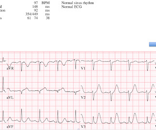
2 middle aged males presented with chestpain. Which had the more severe chestpain at the time of the ECG? Patient 2 at the bottom with a very subtle OMI complained of 10/10 chestpain at the time the ECG was recorded. 414 patients were included in the analysis.
Written by Jesse McLaren A 50 year old presented to triage with one hour of chestpain, and the following ECG labeled normal by the computer (GE Marquette SL) algorithm. Emergent cardiac outcomes in patients with normal electrocardiograms in the emergency department. What do you think? Here is her ECG: What do you think?
A 50-something man presented in shock with severe chestpain. A 12-lead electrocardiogram, lead V4R , and leads V7-9 were recorded on admission. For review — GO TO: The June 4, 2018 post ( LA-LL reversal ). The July 29, 2018 post ( LA-RA reversal ). The November 4, 2018 post ( Leads V1,V2 misplacement ).
Submitted by anonymous, written by Pendell Meyers A woman in her 50s presented to the Emergency Department with chestpain and shortness of breath that woke her from sleep, with diaphoresis. See these other cases of arterial pulse tapping artifact: A 60 year old with chestpain Are these Hyperacute T-waves? 2010.12.162.
Case written and submitted by Ryan Barnicle MD, with edits by Pendell Meyers While vacationing on one of the islands off the northeast coast, a healthy 70ish year old male presented to the island health center for an evaluation of chestpain. The chestpain started about one hour prior to arrival while bike riding.
Written by Jesse McLaren Two 70 year olds had acute chestpain with nausea and shortness of breath, and called paramedics. DIagnostic accuracy oF electrocardiogram for acute coronary OCClusion resulTing in myocardial infarction (DIFOCCULT study). Eur Heart J 2018 4. Who needs the cath lab? Aslanger et al. Lemkes et al.
It is from a 50-something with chestpain: What do you think? Meyers, Weingart and Smith in their 2018 OMI Manifesto. I like to start my ECG assessment in patients with new chestpain by looking for at least 1 or 2 leads that I know are definitely abnormal. This was sent to me by a friend.
Written by Jesse McLaren A 70 year old with prior MIs and stents to LAD and RCA presented to the emergency department with 2 weeks of increasing exertional chestpain radiating to the left arm, associated with nausea. Echo showed new anterior regional wall motion abnormality and decrease EF from 60% to 45%. Clin Cardiol 2022 4.
The finding of dynamic ST-T wave changes on serial tracings in association with a change in chestpain symptoms ( SEE My Comment in the July 21, 2020 post ). Any ST elevation in inferior leads that occurs in association with mirror-image opposite ST depression in lead aVL. ST depression that is maximal in leads V2-to-V4.
She went on to describe her chestpain as a "buffalo sitting on my chest" and a "weird" sensation in her jaw for 1 hour prior to arrival, associated with lightheadedness and diaphoresis. The patient was given fentanyl initially for chestpain with minimal effect and then vomited which was followed by zofran and famotidine.
The best course is to wait until the anatomy is defined by angio, then if proceeding to PCI, add Cangrelor (an IV P2Y12 inhibitor) I sent the ECG and clinical information of a 90-year old with chestpain to Dr. McLaren. All electrocardiograms (ECGs) and coronary angiograms were blindly analyzed by experienced cardiologists.
The patient contacted EMS after a few hours of chestpain that started 5:30 AM. The pain was described as 6/10 radiating to the right shoulder. The chestpain was described as both sharp and pressure like. Below is the post -PCI electrocardiogram. He is otherwise healthy.
Acute chestpain and a bizarre ECG Bizarre (Hyperacute??) Electromechanical association: a subtle electrocardiogram artifact. Incredibly , this case was just published in Circulation on January 22, 2018 (thanks to Brooks Walsh for finding this!) 2018; 137: 402-404. 2018; 137: 402-404. What do you think?
A Deep Neural Network learning algorithm outperforms a conventional algorithm for emergency department electrocardiogram interpretation. But lead V2 has a worrisome amount of ST elevation, and in a chestpain patient, I would be worried about STEMI. I do research on Cardiologs' algorithm: Smith SW et al. What an honor.
male with a history of HTN and ETOH developed squeezing epigastric abdominal pain with associated vomiting and diaphoresis, followed by a syncopal episode which lasted about 10 seconds. When medics arrived, he denied any chestpain, shortness of breath, or palpitations prior to the syncopal episode.
Cardiology felt her chestpain to be, most likely, the result of coronary supply-demand mismatch in the context of HCM endothelial remodeling (i.e. New insights into the use of the 12-lead electrocardiogram for diagnosing acute myocardial infarction in the emergency department. Below are two examples of this. 40; 1234-1241.
Edits by Meyers and Smith A man in his 70s with PMH of hypertension, hyperlipidemia, type 2 diabetes, CVA, dual-chamber Medtronic pacemaker, presented to the ED for evaluation of acute chestpain. Triage ECG: What do you think? This is diagnostic of proximal LAD occlusion. This is a huge anterolateral OMI. I cannot be anything else.
He denied chestpain or shortness of breath. In the clinical context of weakness and fever, without chestpain or shortness of breath, the likelihood of Brugada pattern is obviously much higher. Induced Brugada-type electrocardiogram, a sign for imminent malignant arrhythmias. PM Cardio digitized version.
The utility of the triage electrocardiogram for the detection of ST-segment elevation myocardial infarction. October 2018. We record ECGs in triage on every patient with chestpain, and some other indications, and this amounts to 8000 ECGs in triage each year, costing at most $200,000 (8000 x $20.00).
She denied chestpain and denied feeling any palpitations, even during her triage ECG: What do you think? 1211-1212 CrossRef View Record in Scopus Google Scholar 2 FI Marcus, W Zareba The electrocardiogram in right ventricular cardiomyopathy/dysplasia. J Electrocardiol, 42 (2009), pp.
A middle-aged woman with history of hypertension presented to another hospital approximately 2 hours after onset of chestpain and shortness of breath. Early Continuous ST Segment Monitoring in Unstable Angina: Prognostic Value Additional to the Clinical Characteristics and the Admission Electrocardiogram. mm STE in V1 and 1.5-2.0
It was from a patient with chestpain: Note the obvious Brugada pattern. Induced Brugada-type electrocardiogram, a sign for imminent malignant arrhythmias. The elevated troponin was attributed to either type 2 MI or to non-MI acute myocardial injury. There is no further workup at this time. This patient ruled out for MI.
The patient denied any chestpain whatsoever, and a troponin at zero and 2 hours were both undetectable. Fever not only unmasks a Brugada-type electrocardiogram (ECG) but also increases the risk of ventricular tachyarrhythmias such as ventricular fibrillation (VF) or sudden cardiac death. Heart Rhythm 2018.
We organize all of the trending information in your field so you don't have to. Join thousands of users and stay up to date on the latest articles your peers are reading.
You know about us, now we want to get to know you!
Let's personalize your content
Let's get even more personalized
We recognize your account from another site in our network, please click 'Send Email' below to continue with verifying your account and setting a password.


Let's personalize your content