This site uses cookies to improve your experience. To help us insure we adhere to various privacy regulations, please select your country/region of residence. If you do not select a country, we will assume you are from the United States. Select your Cookie Settings or view our Privacy Policy and Terms of Use.
Cookie Settings
Cookies and similar technologies are used on this website for proper function of the website, for tracking performance analytics and for marketing purposes. We and some of our third-party providers may use cookie data for various purposes. Please review the cookie settings below and choose your preference.
Used for the proper function of the website
Used for monitoring website traffic and interactions
Cookie Settings
Cookies and similar technologies are used on this website for proper function of the website, for tracking performance analytics and for marketing purposes. We and some of our third-party providers may use cookie data for various purposes. Please review the cookie settings below and choose your preference.
Strictly Necessary: Used for the proper function of the website
Performance/Analytics: Used for monitoring website traffic and interactions
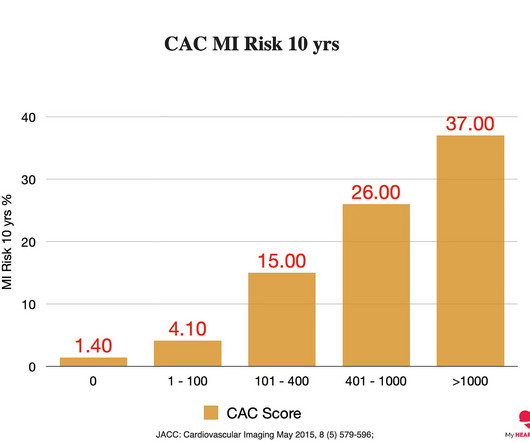
A cardiac CT is a low-dose CT scan of your heart that assesses whether or not you have plaque in your coronary arteries and, if so, how much. In general, the more plaque you have, the higher your risk of a heart attack over the next 10 years. 6, 2017 7 JACC: Cardiovascular Imaging May 2015, 8 (5) 579-596; J Am Coll Cardiol.
This registry will aim to provide world-wide physicians the most accurate information on coronary plaque to improve cardiovascular risk prediction and support the selection of patient-specific treatment,” said Dr. De Cecco. The ultimate goal is to positively impact cardiovascular health globally with a reduction in cardiovascular events."
However, most adults will start to develop advanced plaque in their coronary arteries early in life. By age 66, more than half of all females will have evidence of advanced plaque in their coronary arteries, as seen on a CT calcium score. Coronary atherosclerosis, as evidenced by an abnormal CAC score, is a measure of advanced plaque.
Incident carotid plaques and their vulnerability were detected by carotid ultrasound at follow-up (2021). Higher sdLDL-C or sdLDL-C/LDL-C ratio, but not LDL-C, was significantly associated with an increased risk of incident carotid plaques. years (SD=0.14). years (SD=0.14). 9.90];P=0.027;Pfor linear trend=0.025).
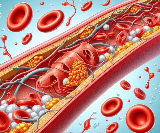
A heart attack is when that plaque ruptures and stops blood flow down the artery. 2017 Apr 12;6(4):e005333. The pathology that causes heart disease (atherosclerosis) is, by definition, the abnormal retention of a cholesterol particle in the artery wall. Cholesterol is an essential part of that process. J Am Heart Assoc.
Carotid ultrasound results were divided into two groups based on the presence or absence of plaque. Carotid plaque was observed in 1140 (43.5%) subjects and CACS>0 in 1172 (44.7%) subjects. Carotid plaque was observed in 1140 (43.5%) subjects and CACS>0 in 1172 (44.7%) subjects. 1692 (64.6%) were male.
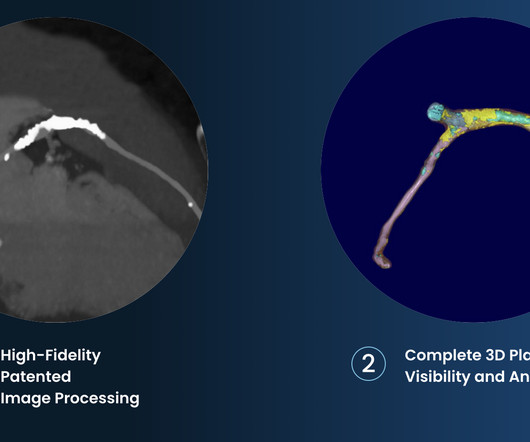
Although carotid plaques can be identified on CT angiography (CTA), interpretation is challenging for frontline physicians. Quantification of plaque volume/composition requires much manual effort. For detection of calcific and hypodense plaque components, respectively, the model achieved sensitivity of 96.5% (95%CI:89.3-99.1%)
people from the general population), coronary artery calcium scores (CACS) are higher, indicating more calcification and the presence of atherosclerotic plaques. Calcified plaques are known to be more stable and less prone to rupture and lead to a heart attack. When comparing athletes to control groups (i.e., hours per week).
The 2017 ESC guidelines on the diagnosis and treatment of peripheral arterial diseases, in collaboration with the European Society for Vascular Surgery (ESVS).” “Diagnosis and treatment of ischemia-producing coronary stenoses improves 5-year survival of patients undergoing major vascular surgery.” Journal of Vascular Surgery, Mar.
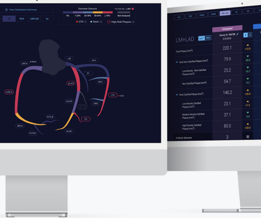
2-7 Through its FDA-cleared solutions driven by artificial intelligence, Cleerly, founded in 2017, supports comprehensive phenotyping of coronary artery disease, as determined from advanced non-invasive CT imaging. With Plaque Features Associated with False Positives. Barcelona, Spain. 4 Chiou A, Hermel M, Miller G et al.
Therefore, if someone presents with an event earlier than this age, they likely have been building up plaque for a considerable period prior to this. A CT CAC score of 0 means a person has no calcified coronary artery plaque and also means their risk of a heart attack over the next ten years is probably less than 2%. N Engl J Med.
2017 Dec 29;7(1):e007664. 5 High intensity interval training induces beneficial effects on coronary atheromatous plaques – a randomized trial, European Journal of Preventive Cardiology , 2022;, zwac309, 6 FOURIER Steering Committee and Investigators. 2017 May 4;376(18):1713-1722. J Am Heart Assoc. J Am Heart Assoc.
His ECG at the accepting facility is shown below: Accepting facility ECG The team reviewed his angiography films with an interventionalist and thought they were suspicious for plaque rupture in LAD, but they were not confident.
Atherosclerotic cardiovascular disease (ASCVD), caused by plaque buildup in arterial walls, is one of the leading causes of disability and death worldwide.1,2 7 Research has shown inflammation plays a significant role in the development of atherosclerosis and ASCVD,8-10 and even the formation of plaque.11 4 In the U.S.
The history of diagnostic testing for coronary disease shows that we are better and better at identifying disease, but simply identifying coronary plaque isn’t the home run people think because of how ubiquitous the development of coronary plaque is in humans.
The cause of angina usually involves inadequate blood flow reaching the heart muscle because of significant narrowing of the artery due to plaque buildup. There are many ways it can present but this is the most common. But coronary stenting is not the only way to reduce symptoms of angina.
Here’s the angiogram of the RCA : No thrombus or plaque rupture in the RCA (or any coronary artery) was found. This MI wasn’t caused by a ruptured plaque of CAD - it was a coronary artery dissection of the RCA. Angiography Angiography was performed after aspirin and heparin were started.
plaque disruption), the T waves still manifest markings of a previous state of suboptimal coronary flow that resolved: Type II supply-demand mismatch in the setting of extreme bradycardia. 2] Although the clinical context in today’s case does not fit these descriptors for Type I OMI (e.g. Chapter 17: Ventricular Arrhythmias. 2] Meyers, H.
The coronary angiogram revealed no critical stenosis, or acute plaque ulceration. Surawicz and Knilans report that intense catecholamine surge, or severe maladjustment of the autonomic nervous system, can manifest “cerebral T waves” in the absence of an acute intracranial process. Furthermore, pertinent electrolyte values (e.g. Josephson, M.
She was treated medically for NonSTEMI, pending next day cath, which showed ulcerated plaque and a 60% thrombotic stenosis in the LAD distal to the first diagonal. A formal contrast echo was done at this point : Normal estimated left ventricular ejection fraction, 65%. Regional wall motion abnormality-distal septum and apex. It was stented.
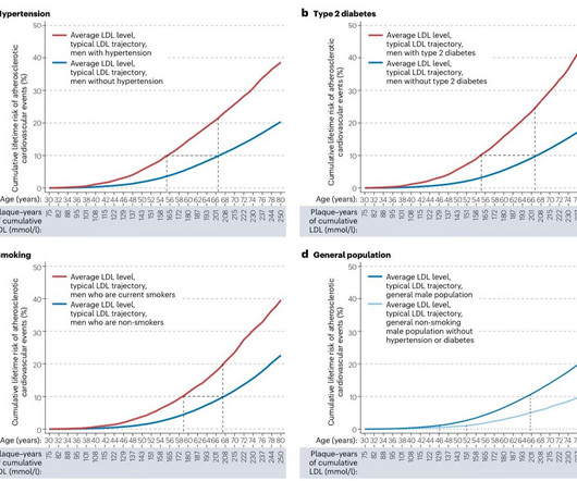
A higher cumulative LDL cholesterol exposure equals a higher likelihood of plaque in the coronary arteries, known as atherosclerosis. A heart attack occurs when plaque in your coronary artery ruptures and causes a clot to form, which stops blood flow to the heart muscle, causing it to die. 2017 Apr;91:1-9. Vascul Pharmacol.
BACKGROUND:A modified computed tomography angiography (CTA)based Carotid Plaque Reporting and Data System (Plaque-RADS) classification was applied to a cohort of patients with embolic stroke of undetermined source to test whether high-risk Plaque-RADS subtypes are more prevalent on the ipsilateral side of stroke.
A 2017 trial named CULPRIT SHOCK found that in patients with cardiogenic shock, a strategy of culprit vessel PCI only was associated with better outcomes than immediate multivessel PCI. As per Dr. Frick use of intravascular imaging ( IVUS or OCT ) could have helped to find plaque rupture and identify the "true" culprit.
We organize all of the trending information in your field so you don't have to. Join thousands of users and stay up to date on the latest articles your peers are reading.
You know about us, now we want to get to know you!
Let's personalize your content
Let's get even more personalized
We recognize your account from another site in our network, please click 'Send Email' below to continue with verifying your account and setting a password.


Let's personalize your content