This site uses cookies to improve your experience. To help us insure we adhere to various privacy regulations, please select your country/region of residence. If you do not select a country, we will assume you are from the United States. Select your Cookie Settings or view our Privacy Policy and Terms of Use.
Cookie Settings
Cookies and similar technologies are used on this website for proper function of the website, for tracking performance analytics and for marketing purposes. We and some of our third-party providers may use cookie data for various purposes. Please review the cookie settings below and choose your preference.
Used for the proper function of the website
Used for monitoring website traffic and interactions
Cookie Settings
Cookies and similar technologies are used on this website for proper function of the website, for tracking performance analytics and for marketing purposes. We and some of our third-party providers may use cookie data for various purposes. Please review the cookie settings below and choose your preference.
Strictly Necessary: Used for the proper function of the website
Performance/Analytics: Used for monitoring website traffic and interactions
mm has been described in normal subjects) Overall impression: In my opinion and experience, this ECG most likely represents a normal baseline ECG, but with a small chance of pericarditis instead. I texted this to Dr. Smith without any information, and this was his reply: "This could be pericarditis but probably is normal variant."
BACKGROUND:Pericardial late gadolinium enhancement (LGE) is usually associated with active pericarditis, but it is not infrequently found in patients after cardiac surgery even a long time after the intervention. All patients were asymptomatic, and no specific treatment for pericarditis was started.
She was diagnosed with pericarditis and spent one day in the hospital without events. Much more classic findings of pericarditis. Learning Points: Pericardial effusion is a key piece of information for the diagnosis and prognosis of pericarditis. Prac 15(17), 2017. Below you will see serial ECGs from that hospitalization.
2017 Oct 1;177(10):1520-1522. In a patient with pericarditis — OR — a large heart on chest X-ray — OR — simply unexplained dyspnea ( as in the November 28, 2022 post) — recognition of electrical alternans should suggest the possibility of a significant pericardial effusion that may be associated with tamponade. J Am Coll Cardiol.
Once they returned in 2017, the team performed open heart surgeries on two patients (Ventricular Septal Defect and Mitral Valve Replacement). Together, they have operated nine Patent Ductus Arteriosus and two Chronic Constrictive Pericarditis between 2012 and 2015.
The second most common cause of medical cardiac tamponade is acute idiopathic pericarditis. Less common etiologies include uremia, bacterial or tubercular pericarditis, chronic idiopathic pericarditis, hemorrhage, and other causes such as autoimmune diseases, radiation, myxedema, etc. 2017 Nov;35(4):525-537. 2013.06.023.
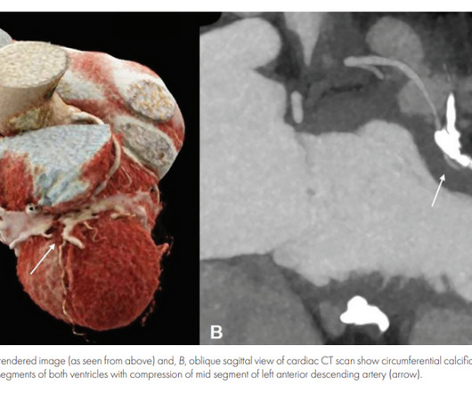
Inflammatory pericarditis can occur in differential fashion. For example, the most common chronic pericarditis tuberculosis affects the fibrinous layer. Post MI pericarditis involves the epicardium. Diastolic Coronary Artery Compression in Constrictive Pericarditis. Angina caused by calcific constrictive pericarditis.
Traditionally used as an anti-inflammatory for pericarditis (inflammation of the lining of the heart), it has recently been shown to result in fewer major heart events in those with a recent heart attack. 2017 Dec 29;7(1):e007664. 2017 May 4;376(18):1713-1722. It is an easy win, frequently missed. J Am Heart Assoc. N Engl J Med.
The initial computer and final cardiology interpretation was a differential: “ST elevation, consider early repolarization, pericarditis, or injury.” J Electrocardiol 2017 2. But STEMI criteria ignore all this and look at ST segments in isolation. McLaren, Meyers, Smith and Chartier.
They include myocardial ischemia, acute pericarditis, pulmonary embolism, external compression due to mass over the right ventricular outflow tract region, and metabolic disorders like hyper or hypokalemia and hypercalcemia. 2017 Mar;110(3):188-195. J Cardiovasc Electrophysiol. 2020 Sep;31(9):2474-2483. Arch Cardiovasc Dis.
The exception is with postinfarction pericarditis , in which a completed transmural infarct results in inflammation of the subepicardial myocardium and STE in the distribution of the infarct, and which results in increased STE and large upright T-waves. These findings together are more commonly seen with pericarditis.
CMAJ 2017 Vassallo SU, Delaney KA, Hoffman RS, et al. Prominent J waves and ventricular fibrillation caused by myocarditis and pericarditis after BNT162b2 mRNA COVID-19 vaccination. Indian Pacing Electrophysiol J 2004 Antzelevitch C, Yan G. J wave syndromes. Heart Rhythm 2010 Hudzik B, Gasior M. J-waves in hypothermia.
We organize all of the trending information in your field so you don't have to. Join thousands of users and stay up to date on the latest articles your peers are reading.
You know about us, now we want to get to know you!
Let's personalize your content
Let's get even more personalized
We recognize your account from another site in our network, please click 'Send Email' below to continue with verifying your account and setting a password.


Let's personalize your content