This site uses cookies to improve your experience. To help us insure we adhere to various privacy regulations, please select your country/region of residence. If you do not select a country, we will assume you are from the United States. Select your Cookie Settings or view our Privacy Policy and Terms of Use.
Cookie Settings
Cookies and similar technologies are used on this website for proper function of the website, for tracking performance analytics and for marketing purposes. We and some of our third-party providers may use cookie data for various purposes. Please review the cookie settings below and choose your preference.
Used for the proper function of the website
Used for monitoring website traffic and interactions
Cookie Settings
Cookies and similar technologies are used on this website for proper function of the website, for tracking performance analytics and for marketing purposes. We and some of our third-party providers may use cookie data for various purposes. Please review the cookie settings below and choose your preference.
Strictly Necessary: Used for the proper function of the website
Performance/Analytics: Used for monitoring website traffic and interactions
This update summarizes relevant clinical data published since the 2017 American Heart Association scientific statement on KD related to diagnosis, cardiac imaging in acute KD treatment, and long-term management. Recent data have advanced the understanding of safety and dosing for several anti-inflammatory therapies in KD.
6 This novel study marks a significant milestone in the field, evaluating the effectiveness of FFR CT in detecting ischemia-producing coronary stenosis in patients with severe PAD. Diagnosis and treatment of ischemia-producing coronary stenoses improves 5-year survival of patients undergoing major vascular surgery.” 2024, [link].
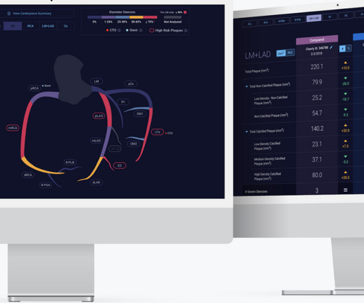
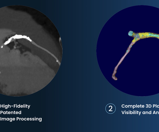
2-7 Through its FDA-cleared solutions driven by artificial intelligence, Cleerly, founded in 2017, supports comprehensive phenotyping of coronary artery disease, as determined from advanced non-invasive CT imaging. High Diagnostic Accuracy Of AI-Ischemia in Comparison To PET, FFR-CT, SPECT, and Invasive FFR: A PACIFIC Sub-Study.
The company is also pursuing an indication for non-invasive measurement of fractional flow reserve (FFR CT ), uniquely derived from its plaque algorithm, to measure coronary blockages and the extent of ischemia. 2017 23, April 2020; Available from: [link]. Cardiovasc. 6 (3) (2019).
or basilar ischemia. 2017 Sep-Oct;50(5):561-569. Epub 2017 Apr 19. CTA head and neck were obtained and showed no evidence of intracranial hemorrhage, large vessel occlusion stroke (what a helpful and apt name for an acute arterial occlusion paradigm, by the way.), EKG on arrival to the ED is shown below: What do you think?
2017 Oct 1;177(10):1520-1522. Alternation in ST segment appearance ( or in the amount of ST elevation or depression ) — is often linked to ischemia. J Am Coll Cardiol. 2006 Jan 17;47(2):269-81. doi: 10.1016/j.jacc.2005.08.066. 2005.08.066. Epub 2006 Jan 4. PMID: 16412847. Moore PK, Raffel KE, Whitman IR. JAMA Intern Med. 2017.3191.
There is no definite evidence of acute ischemia. (ie, Simply stated — t he patient was having recurrent PMVT without Q Tc prolongation, and without evidence of ongoing transmural ischemia. ( Some residual ischemia in the infarct border might still be present. Both episodes are initiated by an "R-on-T" phenomenon.
This was interpreted by the treating clinicians as not showing any evidence of ischemia. This is a critically important determination because of the 2017 AHA/ACC/HRS Guidelines for Management of Patients with Ventricular Arrhythmias and the Prevention of Sudden Cardiac Death. Here is his presenting ECG: ECG 1, t = 0 What do you think?
Learning Point: Concordant ST segment elevation can arise from profound ischemia triggered by ventricular tachycardia (VT), or it may represent an exaggerated basal ST change accompanying tachycardia. The patient rapidly regained consciousness, reporting no residual pain. A peak troponin level of 70 ng/L was observed.
This study compares post-thrombectomy outcomes in both groups, matched by initial NIHSS scores.Method:From October 2017 to March 2023, we studied LVO cases undergoing thrombectomy for acute ischemic stroke. Successful recanalization was defined as Thrombolysis in Cerebral Ischemia score ≥2b. However, clinical outcomes remain similar.
Methods:We retrospectively evaluated AIS patients treated at our institution between January 2017 and January 2023. Modified Treatment in Cerebral Ischemia (mTICI) score was used to determine reperfusion status, with mTICI > 2b considered successful.
A majority (62.5%) of those presenting with ‘normal’ ECGs had the cath lab activated without any ECG being labeled ‘STEMI’ by automated interpretation – based on signs of Occlusion MI including ECG changes, regional wall motion abnormality on bedside ultrasound, or refractory ischemia. 2017 ; 24 ( 1 ): 120 - 124 2. Acad Emerg Med.
For now, the 2017 AHA/ACC/HRS guidelines for asymptomatic patients that have inducible types of Brugada syndrome recommend observation without any specific therapies or interventions [8]. 2017 AHA/ACC/HRS guideline for management of patients with ventricular arrhythmias and the prevention of sudden cardiac death.
There is broad subendocardial ischemia as demonstrated by STE aVR with concomitant STD that almost appears appropriately maximal in Leads II and V5. There is LBBB-like morphology with persistent patterns of subendocardial ischemia. This is the initial ECG: The QRS is widened with a regular cadence, and there are no discernable P waves.
There is normal R-wave progression in the precordial leads with no evidence of ischemia. 21, 2017 ). Here the image quality is enhanced using the PM Cardio app. What do you think? The presenting ECG shows SR with narrow QRS complexes.
mEq/L for Mg++ ) — given that ECF levels of these cations comprise only ~1-2% of total body stores ( Jahnen-Dechent and Ketteler — Clin Kidney J 5(Suppl 1):i3-i14, 2012 — and — Udensi and Tchounwou — Int J Clin Exp Physiol 4(3): 111-122, 2017 ). mEq/L for K+ and 1.76
IntroductionPituitary apoplexy is a serious, emergent, and potentially dangerous condition of the pituitary in which the gland is affected by hemorrhage and/or ischemia typically in the setting of an underlying adenoma. Rutkowski’s 2017 study was the only to assess outcomes prior to versus after intervention at the 72 hours timepoint.
hours ECG: Not much change hs troponin I peaks at 500 ng/L 8 hours Next morning Urine drug screen: Amphetamine, Methamphetamine, Fentanyl, Fentanyl metabolite Formal Bubble Contrast Echocardiogram: Indications for Study: Silent Ischemia. SUMMARY Normal left ventricular cavity size. Normal estimated left ventricular ejection fraction.
The ORBITA trial published in 2017 attempted to answer this question by taking people with significant narrowing of a coronary artery and then randomising them to either medications alone in addition to a sham procedure or medications plus stenting 2. Subscribe now 1 ISCHEMIA Research Group. N Engl J Med. 2020 Apr 9;382(15):1395-1407.
They include myocardial ischemia, acute pericarditis, pulmonary embolism, external compression due to mass over the right ventricular outflow tract region, and metabolic disorders like hyper or hypokalemia and hypercalcemia. 2017 Mar;110(3):188-195. J Cardiovasc Electrophysiol. 2020 Sep;31(9):2474-2483. Arch Cardiovasc Dis.
For now, the 2017 AHA/ACC/HRS guidelines for asymptomatic patients that have inducible types of Brugada syndrome recommend observation without any specific therapies or interventions [8]. 2017 AHA/ACC/HRS guideline for management of patients with ventricular arrhythmias and the prevention of sudden cardiac death.
European Heart Journal 38(41):3082-3089; November 1, 2017. There is no way the ST-T wave should change from lead V4-to-V5 as we see here given the similar all negative QRS appearance in these 2 leads, unless there is acute ischemia. These include disproportionately large upright and negative T waves in leads V2 and V6, respectively.
However, its refined version LMWH, though made it more palatable & user friendly, it un-apologetically took the sting out of regular heparin, made it less efficacious (more glamorous though) LMWH usage is in CAD widespread , it has suspect value* in true ongoing ischemia in any active ACS situation. 2017 Jun 6;69(22):2692-2695.
It’s important to stress the presence of a normal QRS (i.e., Electrocardiographic differentiation of early repolarization from subtle anterior ST-segment elevation myocardial infarction. Annals of Emergency Medicine, 60 (1), 45-56. 2] Driver, B.
This strongly suggests reperfusing RCA ischemia. Troponins, echocardiogram An echocardiogram showed inferobasilar hypokinesis, further supporting a diagnosis of regional ischemia , likely of the area supplied by the RCA. There is also a Q-wave in III. There is also subtle STD in V3-V5. The initial troponin I was elevated at 0.75
12,16 In 2017, CANTOS (Canakinumab Anti-inflammatory Thrombosis Outcomes Study) provided proof-of-principle that inflammation inhibition in the absence of lipid lowering can significantly reduce cardiovascular event rates and helped to define the interleukin-1 (IL-1) to IL-6 to CRP pathway as a central target in CV disease.16
Such findings would normally suggest primary ischemia with concomitant surveillance of coronary occlusion, but these ST/T changes might very well be secondary to the Escape mechanism at hand. Lead V2 shows RR’ QRS configuration, and although ST depression is otherwise expected here, the discordance is a bit excessive. 2] Meyers, H.
BMJ 2017;359:j4788 doi: 10.1136/bmj.j4788 (Published 2017 November 07) Full text link: [link] Abstract Objective To evaluate how selection of patients for high sensitivity cardiac troponin testing affects the diagnosis of myocardial infarction across different healthcare settings. Sandoval Y et al.
2017 Nov;35(4):525-537. Alternation in ST segment appearance ( or in the amount of ST elevation or depression ) — is often linked to ischemia. The above said — Electrical alternans is a nonspecific ECG sign that may also indicate myocardial ischemia, LV dysfunction and/or possibility of any of a number of other precipitating factors.
In most cases, rather, the culprit is gross ischemia due to myocardial infarction, cardiomyopathy, or advanced coronary artery disease. Unfortunately, today’s case is lacking any such diagnostics, thus I cannot say with certainty that the QT interval is, or is not, culpable in arrhythmogenesis. [1] Wolters-Kluwer: Philadelphia, PA. [2]
JACC 69(23):1694-1703; April 4, 2017. S-wave is in V2 = 17 mm S-wave V4 = 9 mm Total = 26 (not greater than 28), so not LVH by the new rule! Nevertheless, it has the look of LVH. Peguero JG et al. Electrocardiographic criteria for the diagnosis of left ventricular Hypertrophy. Still, sensivity was only 62%, with greater than 90% specificity.
Ischemic ST-segment depression maximal in V1-V4 (versus V5-V6) of any amplitude is specific for Occlusion Myocardial Infarction (versus nonocclusive ischemia). Eur Heart J 2017 Driver BE, Shroff GR, Smith SW. JAHA 2022 Grosmaitre P et al. Arch Cardiovasc Dis 2013 Khan AR et al. Emerg Med J 2017;34(2):119–23.
J Electrocardiology 50(5):561-569; September/October 2017. To me, this makes the ECG nearly diagnostic of ischemia, though if it is LAD occlusion, there should be ST depression in III and aVL, so it is a bit confusing. This makes it almost certain that the ST elevation on the first one is due to ischemia. the more accurate.
Even though they were passed the 12 hour mark traditionally associated with reperfusion benefits, ongoing ischemia requires emergent angiogram On assessment, the patient appeared uncomfortable, leaning forward in his chair. ACS with refractory ischemia and electrical instability are indications for emergent cath regardless of the ECG!
However, the mechanisms underlying brain ischemia in DE are not well understood. Background:Dolichoectasia (DE) is characterized by the abnormal dilation and elongation of brain arteries, leading to high morbidity and mortality, commonly presenting as ischemic stroke.
CMAJ 2017 Vassallo SU, Delaney KA, Hoffman RS, et al. Occurrence of “J Waves” in 12-Lead ECG as a Marker of Acute Ischemia and Their Cellular Basis. Occurrence of "J waves" in 12-lead ECG as a marker of acute ischemia and their cellular basis. Heart Rhythm 2010 Hudzik B, Gasior M. J-waves in hypothermia.
Evidence of acute ischemia (may be subtle) vii. ST segment and T wave abnormalities consistent with or possibly related to myocardial ischemia. And these findings come from OESIL , EGSYS , and Sarasin studies: i: Non-sinus rhythm ii: SVT or VT (obviously, and this makes for an abnormal vital sign anyway) iii. Left BBB vi. LVH or RV d.
There is low voltage in the precordium which always makes reading ischemia harder. In ACS, chest pain is the warning sign of ongoing ischemia. Smith : As Willy says, and as we've said many times before, morphine will resolve pain without resolving ischemia. ECG 1 What do you think? To me, this ECG is not diagnostic.
There is ST elevation in 9/12 leads with ST depression only seen in lead aVR>V1 ( ie, virtually the opposite of what is seen with diffuse subendocardial ischemia in which there is diffuse ST depression except for ST elevation in aVR>V1 ).
Denying patients the potential benefit of revascularization just because their symptoms have lasted a certain amount of time shows poor understanding of the pathophysiology of myocardial ischemia. There were no other causes of dyspnea apparent and thus we can assume that myocardial ischemia started 6 days prior.
We organize all of the trending information in your field so you don't have to. Join thousands of users and stay up to date on the latest articles your peers are reading.
You know about us, now we want to get to know you!
Let's personalize your content
Let's get even more personalized
We recognize your account from another site in our network, please click 'Send Email' below to continue with verifying your account and setting a password.


Let's personalize your content