This site uses cookies to improve your experience. To help us insure we adhere to various privacy regulations, please select your country/region of residence. If you do not select a country, we will assume you are from the United States. Select your Cookie Settings or view our Privacy Policy and Terms of Use.
Cookie Settings
Cookies and similar technologies are used on this website for proper function of the website, for tracking performance analytics and for marketing purposes. We and some of our third-party providers may use cookie data for various purposes. Please review the cookie settings below and choose your preference.
Used for the proper function of the website
Used for monitoring website traffic and interactions
Cookie Settings
Cookies and similar technologies are used on this website for proper function of the website, for tracking performance analytics and for marketing purposes. We and some of our third-party providers may use cookie data for various purposes. Please review the cookie settings below and choose your preference.
Strictly Necessary: Used for the proper function of the website
Performance/Analytics: Used for monitoring website traffic and interactions
KEY Point: Knowing that the most commonly overlooked arrhythmia is AFlutter — suggests that the BEST way to avoid missing the diagnosis of AFlutter is simply to THINK of AFlutter whenever you have a regular SVT at a rate close to 150/minute ( in which you do not clearly see upright sinus P waves in lead II ).
Learning points : Takotsubo can lead to cardiac arrest from ventricular arrhythmia. Most Torsades is the result of a pause-dependent effect that predisposes to development of the malignant arrhythmia ( Dohadwala et al — Heart Rhythm Case Rep 3(2):115-119, 2017 ).
Cardiology was consulted, and the note said "no arrhythmias on telemetry or pacer interrogation." This is supported by the PT note which described a palpably irregular pulse with pauses and marked bradycardia. The final cardiology recommendation was to increase fludrocortisone and midodrine.
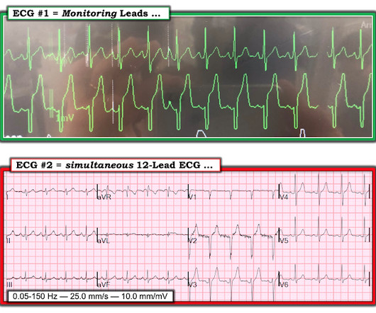
However, he suddenly developed a series of malignant ventricular arrhythmias. Below are printouts of some of the arrhythmias recorded. This time, the arrhythmia did not spontaneously terminate — but rather degenerated to VFib, requiring defibrillation. The arrhythmia starts with a PVC having a short coupling interval.
plaque disruption), the T waves still manifest markings of a previous state of suboptimal coronary flow that resolved: Type II supply-demand mismatch in the setting of extreme bradycardia. Chapter 17: Ventricular Arrhythmias. 2] Although the clinical context in today’s case does not fit these descriptors for Type I OMI (e.g.
Prior to Mizusawa's study, it was thought that the incidence of syncope, arrhythmia, or SCD in this cohort was low [7]. For now, the 2017 AHA/ACC/HRS guidelines for asymptomatic patients that have inducible types of Brugada syndrome recommend observation without any specific therapies or interventions [8]. There was a 0.9%
PVCs N ot generally considered abnormal ECG findings: Isolated PAC, First Degree AV Block, Sinus bradycardia at a rate of 35-45, and Nonspecific ST-T abnormalities (even if different from a previous ECG). Thus, if there is documented sinus bradycardia, and no suspicion of high grade AV block, at the time of the syncope, this is very useful.
This is based on the Sieira et al, 2017, risk calculator , which gives a borderline risk score (2). Regardless of further evaluation, she should avoid bradycardia, AV nodal blockers, Na channel blockers, and fevers. --If Conclusion of this paper: Fever is a great risk factor for arrhythmia events in Brugada Syndrome patients.
We don’t need to think deep, to realize, modalities which take on this arrhythmia head-on has a minuscule role at the population level. Any good physician can easily recognize and manage this arrhythmia with simple measures, advices and liberal use of beta blockers, without the need of tricky drugs like Amiodarone. 2017.11.001.
This ECG shows a sinus bradycardia with a normal conduction pattern (normal PR, normal QRS, and normal QTc), normal axis, normal R-wave progression, normal voltages. Hypothermia can also produce bradycardia and J waves, with a pseudo-STEMI pattern. CMAJ 2017 Vassallo SU, Delaney KA, Hoffman RS, et al. What do you think?
We organize all of the trending information in your field so you don't have to. Join thousands of users and stay up to date on the latest articles your peers are reading.
You know about us, now we want to get to know you!
Let's personalize your content
Let's get even more personalized
We recognize your account from another site in our network, please click 'Send Email' below to continue with verifying your account and setting a password.


Let's personalize your content