This site uses cookies to improve your experience. To help us insure we adhere to various privacy regulations, please select your country/region of residence. If you do not select a country, we will assume you are from the United States. Select your Cookie Settings or view our Privacy Policy and Terms of Use.
Cookie Settings
Cookies and similar technologies are used on this website for proper function of the website, for tracking performance analytics and for marketing purposes. We and some of our third-party providers may use cookie data for various purposes. Please review the cookie settings below and choose your preference.
Used for the proper function of the website
Used for monitoring website traffic and interactions
Cookie Settings
Cookies and similar technologies are used on this website for proper function of the website, for tracking performance analytics and for marketing purposes. We and some of our third-party providers may use cookie data for various purposes. Please review the cookie settings below and choose your preference.
Strictly Necessary: Used for the proper function of the website
Performance/Analytics: Used for monitoring website traffic and interactions
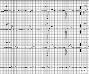
Validation of the Modified Sgarbossa Rule for Diagnosis of STEMI in the Presence of Left Bundle Branch Block. American Heart Journal 170(6):1255-1264; December 2015. Theiling BJ. 0 0 1 41 238 MMRF 1 1 278 14.0 So there is a definite inferior and lateral MI. Here is the first ECG recorded after reperfusion: ST deviation has resolved.
Code STEMI was activated by the ED physician based on the diagnostic ECG for LAD OMI in ventricular paced rhythm. This was several months after the 2022 ACC Guidelines adding modified Sgarbossa criteria as a STEMI equivalent in ventricular paced rhythm). American Heart Journal 170(6):1255-1264; December 2015. Theiling BJ.
Unfortunately, we do not have those images for review, but the operators described a ruptured LAD plaque and they stented this area, which ensures the stability of the plaque. The image on the left shows the LAD before intervention, and the red circled portion on the right indicates the stented region.
This worried the crew of potential acute coronary syndrome and STEMI was activated pre-hospital. As it currently stands, an ST/S ratio >15% should raise awareness for new anterior STEMI. A mid-LAD culprit lesion was identified and stented. Smith comment : V5 and V6 are excessively discordant!!!! References Naidu, S.
He reported a history of ischemic cardiomyopathy with coronary stent placement approximately 10 years prior, but could not recall the specific artery involved. ASA 324mg was administered while a STEMI activation was simultaneously transmitted to the nearest PCI center. A 99% LAD occlusion was stented. Attached is the first ECG.
Dr. Smith illustrates how to measure these parameters with magnified views in his December 21, 2015 post. In the context of the abnormal ST elevation we see in leads III and aVF I interpreted this mirror-image J-point depression as a reciprocal change in this LBBB patient whose ECG is diagnostic of an acute inferior STEMI.
Note: according to the STEMI paradigm these ECGs are easy, but in reality they are difficult. Theres inferior STE which meets STEMI criteria, but this is in the context of tall R waves (18mm) and relatively small T waves, and the STD/TWI in aVL is concordant to the negative QRS. This was false positive STEMI with an ECG mimicking OMI.
He has a history of coronary artery disease and a STEMI two years prior that was treated with primary PCI. At the time of this initial ED ECG, his symptoms were improving ECG #1 on admission to the ED The patient was not seen quickly in the ED as it was a busy shift and the ECG did not meet STEMI criteria. The below ECG was recorded.
He has never been poisoned by the STEMI/NSTEMI paradigm because he has never been to medical school. The Queen of Hearts recognizes this as OMI ("STEMI/STEMI Equivalent"). It was treated with a drug eluting stent. He just graduated from college. He has no medical training, but he has read this blog for years. Lucky Hans.
We organize all of the trending information in your field so you don't have to. Join thousands of users and stay up to date on the latest articles your peers are reading.
You know about us, now we want to get to know you!
Let's personalize your content
Let's get even more personalized
We recognize your account from another site in our network, please click 'Send Email' below to continue with verifying your account and setting a password.


Let's personalize your content