This site uses cookies to improve your experience. To help us insure we adhere to various privacy regulations, please select your country/region of residence. If you do not select a country, we will assume you are from the United States. Select your Cookie Settings or view our Privacy Policy and Terms of Use.
Cookie Settings
Cookies and similar technologies are used on this website for proper function of the website, for tracking performance analytics and for marketing purposes. We and some of our third-party providers may use cookie data for various purposes. Please review the cookie settings below and choose your preference.
Used for the proper function of the website
Used for monitoring website traffic and interactions
Cookie Settings
Cookies and similar technologies are used on this website for proper function of the website, for tracking performance analytics and for marketing purposes. We and some of our third-party providers may use cookie data for various purposes. Please review the cookie settings below and choose your preference.
Strictly Necessary: Used for the proper function of the website
Performance/Analytics: Used for monitoring website traffic and interactions
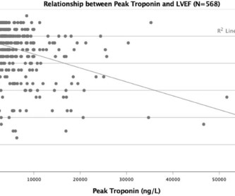
Background The clinical significance of peak troponin levels following ST-elevation myocardial infarction (STEMI) has not been definitively established. Methods A single-centre retrospective observational study was conducted of all patients with STEMI between January 2015 and December 2017. The mean age was 63.6±12
Prompt cath is therefore advised if the post-ROSC shows an acute STEMI. The cause of the abnormal baseline deflections seen in Figure-2 is most likely muscle tremor artifact ( See Bouthillet T — ACLS Med Training, Dec, 2015 ). To Emphasize: The phenomenon of T-QRS-D is not needed in today's case to recognize the acute STEMI.
Saddleback ST Elevation is almost never STEMI 2. An inverted P-wave in lead V2 implies lead misplacement too high Saddleback in STEMI: Here are the only 2 ECGs with V2 "saddleback" that I have ever seen which really represented an LAD Occlusion: Anatomy of a Missed LAD Occlusion (classified as a NonSTEMI) A Very Subtle LAD Occlusion.T-wave
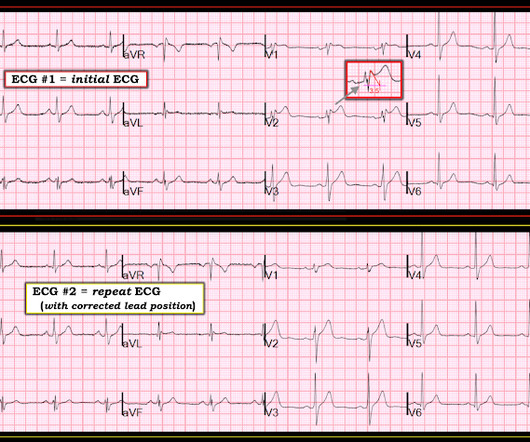
Despite the absence of significant coronary stenosis on her post-arrest cath — the ECG in Figure-1 is clearly diagnostic of an extensive anterolateral STEMI ( presumably from acute LAD [ L eft A nterior D escending ] coronary artery occlusion). The rhythm in ECG #1 is regular and supraventricular at a rate of ~75/minute. What is M INOCA?
Validation of the Modified Sgarbossa Rule for Diagnosis of STEMI in the Presence of Left Bundle Branch Block. American Heart Journal 170(6):1255-1264; December 2015. Theiling BJ. 0 0 1 41 238 MMRF 1 1 278 14.0
In the available view of the sinus rhythm, we see normal variant STE which probably meets STEMI criteria in V4 and V5. In other words, the inferior "ST elevation" is due to the abnormal rhythm, and does not signify OMI or STEMI in any way. This situation has been named "Emery phenomenon." YOU TOO CAN HAVE THE PM Cardio AI BOT!!
The precordial ST-depression pattern on this ECG (and in this clinical setting) should immediately raise suspicion of Posterior STEMI! Posterior STEMI occurs in approximately 15-20% of acute MI, but the vast majority of the time it is seen in conjunction with inferior (Infero-Posterior) or lateral (Postero-Lateral) STEMI (1).
Barely any STE, and thus not meeting STEMI criteria. Annals of Emergency Medicine Cardiology was called to evaluate the patient immediately for emergent cath, but they stated that the ECG did not meet STEMI criteria and elected to wait for further information before proceeding with cath. He was given 6mg IV morphine for ongoing pain.
Code STEMI was activated by the ED physician based on the diagnostic ECG for LAD OMI in ventricular paced rhythm. This was several months after the 2022 ACC Guidelines adding modified Sgarbossa criteria as a STEMI equivalent in ventricular paced rhythm). American Heart Journal 170(6):1255-1264; December 2015. Theiling BJ.
Other trials that evaluated this subject were the WOEST trial (2013), Pioneer AF-PCI trial (2016), and ISAR-TRIPLE (2015). Explanation: The EKG illustrates an inferior STEMI. Explanation: The RE-DUAL PCI trial evaluated differences between triple therapy and dual therapy using antiplatelets/anticoagulant. Incorrect Answers: A and E.
This worried the crew of potential acute coronary syndrome and STEMI was activated pre-hospital. As it currently stands, an ST/S ratio >15% should raise awareness for new anterior STEMI. Smith comment : V5 and V6 are excessively discordant!!!! Here are two examples of HATW’s in the setting of confirmed LVH. References Naidu, S.
From My Comment in the November 15, 2023 post in Dr. Smith's ECG Blog: Clinical Points about MINOCA: Given the literature citing a 5-15% estimated incidence of MINOCA in patients initially diagosed as having a STEMI or "NSTEMI" — it is important to be aware of the more common entities associated with this entity ( See Figure-2 ).
Dr. Smith illustrates how to measure these parameters with magnified views in his December 21, 2015 post. In the context of the abnormal ST elevation we see in leads III and aVF I interpreted this mirror-image J-point depression as a reciprocal change in this LBBB patient whose ECG is diagnostic of an acute inferior STEMI.
The HEART and EDACS scores are helpful to risk stratify patients with chest pain, but they hinge on accurate ECG interpretation: a low score doesn’t apply if the ECG shows STEMI(+)OMI, and shouldn’t be used for STEMI(-)OMI or OMI reperfusion either 2. Lancet 2015 6. Learn the signs of reperfusion: as Dr. Am J Med 2021 5.
Clinical Course The paramedic activated a “Code STEMI” alert and transported the patient nearly 50 miles to the closest tertiary medical center. 2 The astute paramedic recognized this possibility and announced a CODE STEMI. 2015 Oct; 66(4):355-362. Look at the aortic outflow tract. What do you see? Annals of Emergency Medicine.
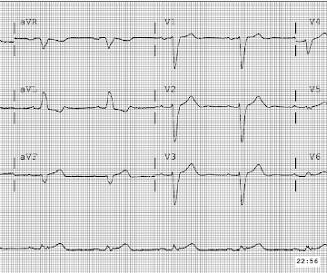
Figure 1-1 My colleague, a faithful student of ECG interpretation, handed me the tracing and said that it warranted STEMI activation because of apparent terminal QRS distortion (TQRSD) in V2. ASA 324mg was administered while a STEMI activation was simultaneously transmitted to the nearest PCI center. Attached is the first ECG.
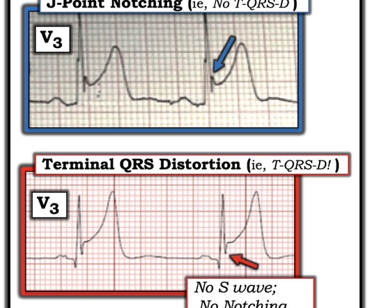
Figure-2: Comparison between ST elevation in lead V3 due to a repolarization variant ( TOP from the 4/27/2019 post) vs acute OMI ( BOTTOM from the 9/20/2015 post) , which manifests T-QRS-D ( See text ). MORE PRACTICE = 4 examples ( 3 positive; 1 not for T-QRS-D ). What About the ECGs shown in Today's Case?
Learning Points: Ectopic atrial rhythm can produce atrial repolarization findings that can be confused for acute ischemia, STEMI, or OMI. To the left of these tracings is schematic illustration of the Emery Phenomenon ( adapted from the 2015 post by Dr. Bojana Uzelac on Armel Carmona’s ECG Rhythms website ).
Anterior STEMI? T-wave inversions and dynamic ST elevation Tachycardia, hyperthyroid, and ST elevation. What is it? 2 Cases of Resolved Chest Pain with Dynamic Terminal T-wave Inversion Is it Wellens' Syndrome? Activate the Cath Lab?
Note: according to the STEMI paradigm these ECGs are easy, but in reality they are difficult. Theres inferior STE which meets STEMI criteria, but this is in the context of tall R waves (18mm) and relatively small T waves, and the STD/TWI in aVL is concordant to the negative QRS. This was false positive STEMI with an ECG mimicking OMI.
He has a history of coronary artery disease and a STEMI two years prior that was treated with primary PCI. At the time of this initial ED ECG, his symptoms were improving ECG #1 on admission to the ED The patient was not seen quickly in the ED as it was a busy shift and the ECG did not meet STEMI criteria. The below ECG was recorded.
Supply-demand mismatch can cause ST Elevation (Type 2 STEMI). Also see these posts of Type II STEMI. An EKG from a year prior was available for comparison: The ED physician noted Initial EKG here read by the computer as a STEMI, however, there is a very poor baseline and a lot of artifact. See reference and discussion below.
He has never been poisoned by the STEMI/NSTEMI paradigm because he has never been to medical school. The Queen of Hearts recognizes this as OMI ("STEMI/STEMI Equivalent"). He just graduated from college. He has no medical training, but he has read this blog for years. He is an ECG tech who hopes to go to medical school.
We organize all of the trending information in your field so you don't have to. Join thousands of users and stay up to date on the latest articles your peers are reading.
You know about us, now we want to get to know you!
Let's personalize your content
Let's get even more personalized
We recognize your account from another site in our network, please click 'Send Email' below to continue with verifying your account and setting a password.


Let's personalize your content