This site uses cookies to improve your experience. To help us insure we adhere to various privacy regulations, please select your country/region of residence. If you do not select a country, we will assume you are from the United States. Select your Cookie Settings or view our Privacy Policy and Terms of Use.
Cookie Settings
Cookies and similar technologies are used on this website for proper function of the website, for tracking performance analytics and for marketing purposes. We and some of our third-party providers may use cookie data for various purposes. Please review the cookie settings below and choose your preference.
Used for the proper function of the website
Used for monitoring website traffic and interactions
Cookie Settings
Cookies and similar technologies are used on this website for proper function of the website, for tracking performance analytics and for marketing purposes. We and some of our third-party providers may use cookie data for various purposes. Please review the cookie settings below and choose your preference.
Strictly Necessary: Used for the proper function of the website
Performance/Analytics: Used for monitoring website traffic and interactions
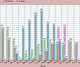
males), referred for a stress echocardiogram (SE), who underwent ESE between July 2020 (immediate post lockdown) and January 2021 according to national safety guidelines, in addition to patients wearing masks during ESE. Methods and results Baseline data were collected prospectively on 740 consecutive patients (mean age 61.4
The study population included a random sample of individuals with HF diagnostic codes (HF with reduced ejection fraction (HFrEF), HF with preserved ejection fraction (HFpEF) and non-specific HF) selected from all participants registered in the THIN database between 1 January 2015 and 30 September 2017.
Hopefully a repeat echocardiogram will be performed outpatient. 2015, March 1). Systolic function normal by visual assessment only, unable to visualize well for further characterization. 1900: RBBB and LAFB are almost fully resolved. 2300: QRS now within normal limits. No other significant injuries were found. No cardiac MRI was done.
The emergent echocardiogram showed normal EF, no WMA, and normal valve function. Figure-1: Illustration of the Emery Phenomenon ( adapted from My Comment in the February 23, 2023 post in Dr. Smith's ECG Blog — and — from the 2015 post by Dr. Bojana Uzelac on Armel Carmona’s ECG Rhythms website ).
His echocardiogram showed normal wall motion. Figure-3: Comparison between ST elevation in lead V3 due to a repolarization variant ( TOP — from 4/27/2019 ) — vs acute OMI ( BOTTOM — from 9/20/2015 ) , which manifests T-QRS-D ( See text ). The patient did well afterward without any recurrence of symptoms.
Due to limitations of echocardiogram in evaluating the right ventricle, magnetic resonance imaging study of the right ventricle along with that of the left ventricle has been reported. 2015 Apr;16(4):353. Effect of exercise on right ventricle. Twenty-one male endurance athletes were compared with untrained control subjects.
A transthoracic echocardiogram showed an LV EF of less than 15%, critically severe aortic stenosis , severe LVH , and a small LV cavity. 2015 Oct; 66(4):355-362. Aortic angiogram did not reveal aortic dissection. The patient was transported to the CCU for further medical optimization where a pulmonary artery catheter was placed.
Cardiac enzymes, CTs, echocardiograms, carotid ultrasounds, and electroencephalography all affected diagnosis or management in Postural blood pressure , performed in only 38% of episodes, had the highest yield with respect to affecting diagnosis (18-26%) or management (25-30%) and determining etiology of the syncopal episode (15-21%).
More troponin values were measured at the cardiac center: 2327- 267 ng/L 0821- 355 ng/L 1108- 305 ng/L An echocardiogram on day three of the patients admission showed an ejection fraction of 46% with abnormal basal inferior and basal lateral segments, and severe aortic stenosis.
We organize all of the trending information in your field so you don't have to. Join thousands of users and stay up to date on the latest articles your peers are reading.
You know about us, now we want to get to know you!
Let's personalize your content
Let's get even more personalized
We recognize your account from another site in our network, please click 'Send Email' below to continue with verifying your account and setting a password.


Let's personalize your content