This site uses cookies to improve your experience. To help us insure we adhere to various privacy regulations, please select your country/region of residence. If you do not select a country, we will assume you are from the United States. Select your Cookie Settings or view our Privacy Policy and Terms of Use.
Cookie Settings
Cookies and similar technologies are used on this website for proper function of the website, for tracking performance analytics and for marketing purposes. We and some of our third-party providers may use cookie data for various purposes. Please review the cookie settings below and choose your preference.
Used for the proper function of the website
Used for monitoring website traffic and interactions
Cookie Settings
Cookies and similar technologies are used on this website for proper function of the website, for tracking performance analytics and for marketing purposes. We and some of our third-party providers may use cookie data for various purposes. Please review the cookie settings below and choose your preference.
Strictly Necessary: Used for the proper function of the website
Performance/Analytics: Used for monitoring website traffic and interactions
The amount of plaque in your coronary arteries can be estimated by looking directly at your coronary arteries with a cardiac CT and calculating your CAC score. This also means that if you have a CAC score of 0, you have no calcified plaque in your coronary arteries. 2 JACC: Cardiovascular Imaging May 2015, 8 (5) 579-596;
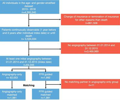
Patients undergoing coronary angiography between January 2014 and December 2015 were included in the analysis. Eligible patients had at least one inpatient coronaryangiogram for suspected coronary artery disease between January 2014 and December 2015.
Ct coronaryangiogram showed normal coronary arteries. Smith note: I think CT coronaryangiogram is reasonable with the elevated troponins and symptoms. He was given aspirin and heparin and transferred to the local cardiac center for further evaluation. He was diagnosed with mild AKI which resolved.
Cardiology felt her chest pain to be, most likely, the result of coronary supply-demand mismatch in the context of HCM endothelial remodeling (i.e. Type II MI), however decided to pursue coronaryangiogram out of an abundance of caution. A mid-LAD culprit lesion was identified and stented. References Naidu, S. Tower-Rader, A.
The diagnostic coronaryangiogram identified only minimal coronary artery disease, but there was a severely calcified, ‘immobile’ aortic valve. Aortic angiogram did not reveal aortic dissection. 2015 Oct; 66(4):355-362. The patient was brought directly to the cardiac catheterization lab for PCI, bypassing the ED.
Case Continued The patient was discharged from the hospital with a plan for a scheduled coronaryangiogram to assess the coronary arteries and the possibility of aortic valve replacement. The vast majority of ischemia from supply demand mismatch is diffuse ST depression, with ST Elevation in aVR.
We organize all of the trending information in your field so you don't have to. Join thousands of users and stay up to date on the latest articles your peers are reading.
You know about us, now we want to get to know you!
Let's personalize your content
Let's get even more personalized
We recognize your account from another site in our network, please click 'Send Email' below to continue with verifying your account and setting a password.


Let's personalize your content