Remove 2015 Remove Cardiac Arrest Remove Chest Pain
article thumbnail

ECG Blog #415 — The Cath showed NO Occlusion!

Ken Grauer, MD

Shortly after arrival in the ED ( E mergency D epartment ) — she suffered a cardiac arrest. BUT — Cardiac catheterization done a little later did not reveal any significant stenosis. Figure-1: The initial ECG in today's case — obtained after successful resuscitation from cardiac arrest. ( No CP ( C hest P ain ).

Blog 155
article thumbnail

ECG Blog #434 — WHY Did this Patient Arrest?

Ken Grauer, MD

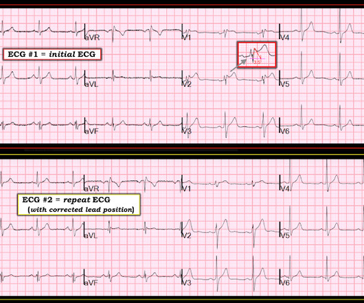
The ECG in Figure-1 — was obtained from a middle-aged man who presented to the ED ( E mergency D epartment ) in cardiac arrest. The cause of the abnormal baseline deflections seen in Figure-2 is most likely muscle tremor artifact ( See Bouthillet T — ACLS Med Training, Dec, 2015 ). Should you activate the cath lab?

Blog 135
article thumbnail

Saddleback ST Elevation. Is it STEMI? Is it type II Brugada?

Dr. Smith's ECG Blog

A 50-something presented with epigastric and chest pain. Epigastric pain, Syncope, and Saddleback ST Elevation A 65 Year Old Man with Chest pain and Precordial ST Elevation Non-Vagal Syncope and Saddleback Morphology in V2 Is this STEMI? Here is his ECG: What do you think? QTc 388 ms. wave in V1??

STEMI 52
article thumbnail

Hypothermia at 18 Celsius in V Fib arrest: CPR, then ECMO rewarming, for 3 hours, then Defib with ROSC. Interpret the ECG.

Dr. Smith's ECG Blog

Although in the context of chest pain such ST depression would be all but diagnostic of posterior OMI, one should make no conclusions in such an unusual case. This Transesophageal ED Echo was recorded: Cardiac POCUS.mov from Stephen Smith on Vimeo. In all leads, there is a 2nd wave after the initial QRS. This is an Osborn wave.