This site uses cookies to improve your experience. To help us insure we adhere to various privacy regulations, please select your country/region of residence. If you do not select a country, we will assume you are from the United States. Select your Cookie Settings or view our Privacy Policy and Terms of Use.
Cookie Settings
Cookies and similar technologies are used on this website for proper function of the website, for tracking performance analytics and for marketing purposes. We and some of our third-party providers may use cookie data for various purposes. Please review the cookie settings below and choose your preference.
Used for the proper function of the website
Used for monitoring website traffic and interactions
Cookie Settings
Cookies and similar technologies are used on this website for proper function of the website, for tracking performance analytics and for marketing purposes. We and some of our third-party providers may use cookie data for various purposes. Please review the cookie settings below and choose your preference.
Strictly Necessary: Used for the proper function of the website
Performance/Analytics: Used for monitoring website traffic and interactions
Even if we stopped here — We could conclude the following: There is marked bradycardia in today's rhythm ( ie, Heart rate in the low 30s ). Finally — If today's patient does not have significant underlying coronary disease — then her bradycardia with AV block may be the result of SSS ( S ick S inus S yndrome ).
Looking first at the long-lead II rhythm strip — there is significant bradycardia , with a heart R ate just under 40/minute. But the point to emphasize — is that it should only take seconds to recognize that there is bradycardia from significant AV block. = Would you approve her for a nonemergent surgical procedure?
That said — obvious findings include: i ) Marked bradycardia! — Section 2F ( 6 pages = the " short " Answer ) from my ECG-2014 Pocket Brain book provides quick written review of the AV Blocks. The rhythm in Figure-1 is complex — and defies precise interpretation without careful study. be regular! —
Use of drugs producing bradycardia like beta blockers in stages III and IV may precipitate low output state. 2014 May-Jun;66(3):392-3. In stage IV, this restrictive filling pattern remains fixed even during Valsalva maneuver. Initial stages (I to III) are considered reversible with treatment. Stage IV is considered as advanced.
Whatever today's rhythm turns out to be — the "good news" is that the bradycardia and degree of AV block is likely to improve as soon as there is reperfusion of the "culprit" artery ( Therefore need for prompt cath with PCI ).
CMAJ 2014. Int J Cardiol 2014. == MY Comment by K EN G RAUER, MD ( 10/13/2022 ): == I suspect most cases of acute OMI that occur in association with WPW — are overlooked! Electrocardiography in the patient with the Wolff-Parkinson-White syndrome: diagnostic and initial therapeutic issues. Am J Emerg Med 1999. Chang and Liu.
Sinus bradycardia, normal conduction, normal axis, normal R wave progression, no hypertrophy. 2014 AHA/ACC guideline for the management of patients with non-ST-elevation acute coronary syndromes: a report of the American College of Cardiology/American Heart Association Task Force on Practice Guidelines. What do you think?
Theres sinus bradycardia, borderline PR interval, narrow QRS; normal axis/R wave progression; low precordial voltages, and subtle peaked T waves (most obvious in V2, but all T waves are symmetric with a narrow base). Theres no prior ECG to compare - but the bradycardia, prolonged PR and peaked T waves could all be from hyperkalemia.
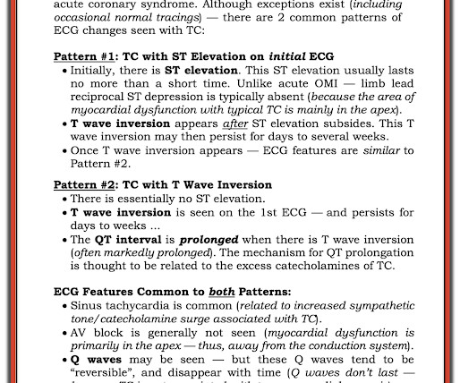
MY Thoughts on ECG #1: The rhythm is sinus bradycardia at a rate just under 60/minute. By way of historical perspective on Takotsubo CM — I've added these 2 pages that I've excerpted from my 2014 ECG-Pocket Brain ePub. Figure-7: Brief review of clinical features of Takotsubo Cardiomyopathy ( Excerpted from Grauer K — ECG-2014-ePub ).
Within ten minutes, she developed bradycardia, hypotension, and ST changes on monitor. Bradycardia and heart block are very common in RCA OMI. Note characteristic ballooning of the apex and hypercontractility of the base during cardiac cath ( Figure excerpted from Grauer K: ECG-2014- Expanded ePub, KG/EKG Press ). =
We organize all of the trending information in your field so you don't have to. Join thousands of users and stay up to date on the latest articles your peers are reading.
You know about us, now we want to get to know you!
Let's personalize your content
Let's get even more personalized
We recognize your account from another site in our network, please click 'Send Email' below to continue with verifying your account and setting a password.


Let's personalize your content