This site uses cookies to improve your experience. To help us insure we adhere to various privacy regulations, please select your country/region of residence. If you do not select a country, we will assume you are from the United States. Select your Cookie Settings or view our Privacy Policy and Terms of Use.
Cookie Settings
Cookies and similar technologies are used on this website for proper function of the website, for tracking performance analytics and for marketing purposes. We and some of our third-party providers may use cookie data for various purposes. Please review the cookie settings below and choose your preference.
Used for the proper function of the website
Used for monitoring website traffic and interactions
Cookie Settings
Cookies and similar technologies are used on this website for proper function of the website, for tracking performance analytics and for marketing purposes. We and some of our third-party providers may use cookie data for various purposes. Please review the cookie settings below and choose your preference.
Strictly Necessary: Used for the proper function of the website
Performance/Analytics: Used for monitoring website traffic and interactions
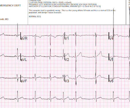
Written by Pendell Meyers A man in his early 40s experienced acute onset chestpain. The chestpain started about 24 hours ago, but there was no detailed information available about whether his pain had come and gone, or what prompted him to be evaluated 24 hours after onset.
A 50-something male who is healthy and active with no previous medical history presented with 5 hours of continuous worrisome chestpain. Chestpain with New LBBB: It helps to actually measure the ST/S ratio A Fascinating Demonstration of ST/S Ratio in LBBB and Resolving LAD Ischemia The cath lab was activated.
Written by Jesse McLaren, with comments from Smith and Grauer A 60 year old presented with three weeks of intermittent non-exertional chestpain without associated symptoms. A prospective validation of the HEART score for chestpain patients at the emergency department. Int J Cardiol 2013 2. Shin YS, Ahn S, Kim YJ.
These were texted to me only with "chestpain." It helps to know that the patient has active chestpain, as Wellen's is a post occlusion (reperfusion) state, with open artery and pain-free. First: 2nd: What was my response? Smith: Young thin black male. Texter: Can't fool you. It was indeed.
A 70-year-old man calls 911 after experiencing sudden, severe chestpain. Acute myocardial infarction with isolated ST-segment elevation in posterior chest leads V7-V9: "hidden" ST-elevations revealing acute posterior infarction. This case comes from Sam Ghali ( @EM_RESUS ). Thanks, Sam! J Am Coll Card 1999; 34:748-753.
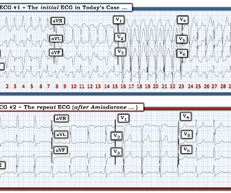
== MY Comment by K EN G RAUER, MD ( 8/22/2020 ): == The patient is a mid-50s man who presented to the ED for new-onset chestpain of ~1 hour duration. He was still having chestpain in the ED at the time ECG #1 was done ( Figure-1 ). His symptoms awakened him from sleep. The Case Continues: Initial troponin was normal.
This was my thought: if this patient presented to the ED with chestpain, then this is an LAD occlusion. Usefulness of automated serial 12-lead ECG monitoring during the initial emergency department evaluation of patients with chestpain. For clarity in Figure-1 — I've labeled the initial ECG in this June 18, 2013 post.
A 40-something male with no previous cardiac disease presented with chestpain. The pain continued and the first high sensitivity troponin I returned at 105 ng/L Another ECG was recorded: The ST segment in aVF has flattened a bit, revealing that there is some STD in addition to the non-specific findings in III and aVL.
As discussed in detail in ECG Blog #228 — this seemingly qualifies as a “ Silent ” MI ( Approximately half of those MIs not accompanied by CP — have some other associated symptom such as syncope, which substitutes as a “chestpain equivalent” ).
No chestpain. In a previously healthy adolescent ( who is 15 years old in today's case ) — the presentation of an acute febrile illness that is without a complaint of chestpain, is highly unlikely to be due to an acute MI. He was hemodynamically stable. How would YOU interpret the ECG in Figure-1 ?
Although this " Imbalance " of precordial T waves is not see n very often — in the “right” clinical setting, it has been associated with recent OMI ( O cclusion-based MI ) , most often from a LCx culprit artery ( See Manno et al: JACC 1:1213, 1983 — and the July 17, 2013 post by Salim Rezaie in ALiEM — and ECG Blog #350 ).
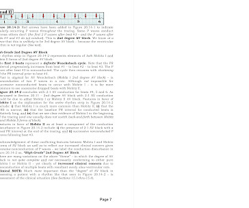
She was hemodynamically stable — and did not have chestpain, lightheadedness or syncope. Section 20 ( 54 pages = the " long " Answer ) from my ACLS-2013-Arrhythmias Expanded Version provides detailed discussion of WHAT the AV Blocks are — and what they are not ! QUESTIONS: HOW would you interpret the rhythm in Figure-1 ?
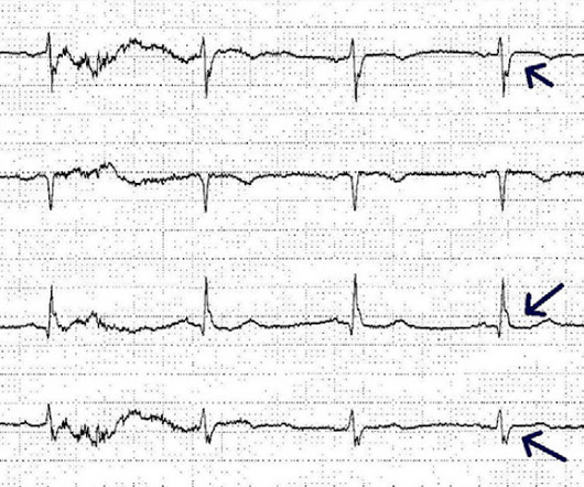
I have enclosed the ECG from a 50-something year old male who complained of chestpain. Steffen wrote: " I remembered the ECG from your blog titled: "STEMI Seen Best in PVC, Diagnosed by Medic, Ignored by Physician" from 2013. I was on scene at his working place (in Germany physicians are on the Emergency vehicles).
and , ii ) That we need to carefully inquire about recent chestpain, paying special attention to the rest of this 12-lead ECG ( looking carefully for signs of acute or recent infarction — because AV Wenckebach is common with acute inferior MI ).
This 50-something otherwise healthy male presented with one hour of epigastric and lower chestpain. One of our fine interns, Daniel Lee, who is also an ECG whiz, found this paper from 2013 and brought it to my attention: The delayed activation wave in non-ST-elevation myocardial infarction.
A 70-something female with no previous cardiac history presented with acute chestpain. She awoke from sleep last night around 4:45 AM (3 hours prior to arrival) with pain that originated in her mid back. She stated the pain was achy/crampy. Over the course of the next hour, this pain turned into a pressure in her chest.
link] A 62 year old man with a history of hypertension, type 2 diabetes mellitus, and carotid artery stenosis called 911 at 9:30 in the morning with complaint of chestpain. He described it as "10/10" intensity, radiating across his chest from right to left. This is written by Willy Frick, an amazing cardiology fellow in St.
The ECG in Figure-1 was obtained from an older woman — who presented with chestpain and palpitations over the previous hour. She had a history of hypertension, and was on medication for this — but she was otherwise healthy. BP = 140/90 mm Hg in association with the rhythm in Figure-1. How would YOU interpret the rhythm in Figure-1 ?
Written by Pendell Meyers, few edits by Smith A man in his 60s with history of stroke and hypertension but no known heart disease presented with chestpain that started on the morning of presentation at around 8am. Here is his triage ECG when he presented at 1657: What do you think?
P.S.: Note that in today's case — the brief description of the chestpain history in this teenager does not sound convincing for an acute cardiac event. But eliciting both a personal and family History on such patients ( inquiring about syncope-presyncope, or malignant arrhythmia symptoms ) — is essential for optimal management.
The best course is to wait until the anatomy is defined by angio, then if proceeding to PCI, add Cangrelor (an IV P2Y12 inhibitor) I sent the ECG and clinical information of a 90-year old with chestpain to Dr. McLaren. His response: “subendocardial ischemia.
Given the history of dyspnea on exertion over a several week period ( but no mention of chestpain ) — and — the finding of deep, symmetric T wave inversion in the anterior leads ( as per Pearl #2 ) — it is possible that the onset of her symptoms is the result of a "Silent MI" ( See ECG Blog #228 for more on "Silent" MI ).
Although this " Imbalance " of precordial T waves is not seen very often — in the “right” clinical setting, it has been associated with recent OMI ( O cclusion-based MI ) from a LCx culprit artery ( See Manno et al: JACC 1:1213, 1983 — and the July 17, 2013 post by Salim Rezaie in ALiEM — and ECG Blog #350 ).
This " imbalance of precordial T waves" is not seen very often — and in the “right” clinical setting, has been associated with recent OMI from a LCx culprit artery ( See Manno et al: JACC 1:1213, 1983 — and the July 17, 2013 post by Salim Rezaie in ALiEM ).
Arch Cardiovasc Dis 2013 Khan AR et al. Smith : this proves my impression that the inferior T-waves on the first ECG are hyperacute. JAHA 2022 Grosmaitre P et al. Significance of atypical symptoms for the diagnosis and management of myocardial infarction in elderly patients admitted to emergency departments.
It was edited by Smith CASE : A 52-year-old male with a past medical history of hypertension and COPD summoned EMS with complaints of chestpain, weakness and nausea. N Engl J Med 2003; 348:1756-1763, 5/1/2013. This was contributed by some folks at Wake Forest: Jason Stopyra, Shannon Mumma, Sean O'Rourke, and Brian Hiestand.
Given her reported chestpain, shortness of breath, and syncope, an ECG was quickly obtained: What do you think? 2013 Sep;26(9):965-1012.e15. She was noted to be tachycardic and her heart sounds were distant on physical exam. She had a normal respiratory effort, and her lungs were clear to auscultation bilaterally. 2013.06.023.
The patient presented with chestpain. I was taught that the tell-tale sign of ischemia vs an electrical abnormality was in the hx, i.e. chestpain for the ischemia and potential syncope for brugada. Only 5-18% of ED patients with chestpain have a myocardial infarction of any kind. Is it Brugada pattern?
Edits by Meyers and Smith A man in his 70s with PMH of hypertension, hyperlipidemia, type 2 diabetes, CVA, dual-chamber Medtronic pacemaker, presented to the ED for evaluation of acute chestpain. Triage ECG: What do you think? This is diagnostic of proximal LAD occlusion. This is a huge anterolateral OMI. I cannot be anything else.
He denied chestpain or shortness of breath. In the clinical context of weakness and fever, without chestpain or shortness of breath, the likelihood of Brugada pattern is obviously much higher. Today's patient presented with acute weakness, syncope and fever, but no chestpain or shortness of breath.
Other trials that evaluated this subject were the WOEST trial (2013), Pioneer AF-PCI trial (2016), and ISAR-TRIPLE (2015). ACS QID 3103 A 64 year old Caucasian male with a history of extensive tobacco use, hypertension, hyperlipidemia, and obesity presents with acute onset chestpain. Incorrect Answers: A and E. Question 2.
He denied any chestpain or shortness of breath and stated he felt at his baseline yesterday prior to drug use. They recommended repeating his ECG and awaiting troponin since the patient did not have any chestpain. He complained of generalized weakness and left lower extremity numbness. What is it?
His comments/questions are inserted below the ECG: A 50-something woman presented with 3 days of intermittent chestpain that became worse on the day of presentation, with diaphoresis and radiation to the left arm, as well as abdominal pain. American heart journal 2010;160:995-1003,e1-8. O'Gara PT, Kushner FG, Ascheim DD, et al.
It was from a patient with chestpain: Note the obvious Brugada pattern. This definition was changed following an expert consensus panel in 2013 — so that at the present time, all that is needed to diagnose Brugada Syndrome is a spontaneous or induced Brugada-1 ECG pattern, without need for additional criteria.
Am J Cardiol 12(9):1379-1383; Nov 2013. the optimum QT correction formula for patients with chestpain was found to be unique for each individual ; it is a correction factor that can be calculated real-time for each patient by taking multiple measurements over a range of heart rates. Musat DL et al.
A 26 year old male presented with syncope and chestpain. No signs of OMI" The chestpain resolved after some time, and another ECG was recorded: The ST Elevation is nearly gone. Syncope was sudden and without prodrome, and resulted in head trauma with a scalp laceration. Smith : I recognize this as a STEMI mimic.
A man in his early 30s was walking when he developed central chestpain which was non-radiating, then had a syncopal event with bowel incontinence, and when he woke up he had ongoing chestpain. Notes never having symptoms like this before, pain is so severe its causing SOB. He called 911.
The ECG in Figure-1 was obtained from a middle-aged man who presents to the ED ( E mergency D epartment ) with 6 hours of chestpain. Figure-1: The initial ECG in today's case obtained from a middle-aged man with 6 hours of chestpain. ( He is hemodynamically stable.
A middle-age woman with no previous cardiac history called 911 for chestpain. This was her prehospital ECG: What do you think? There is sinus rhythm with RBBB and obvious LAD OMI (proximal LAD occlusion): hyperacute T-waves in I, aVL and minimal STE in V1, V2.
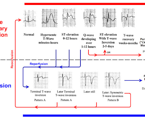
Scenario 1 : The patient presents with 24 hours of substernal chestpain. Ninety percent of patients with reperfusion attained a maximum T wave negativity of 3 mm or more within 48 hours after the onset of chestpain in the lead that initially displayed the greatest ST segment elevation. Below is his presentation ECG.
He had no chestpain, dyspnea, or any other anginal equivalent, and his vital signs were normal. The cardiologist thought she had stent thrombosis which is possible, but I do not necessarily think is sufficient to explain her complete hemodynamic collapse. SanzRuiz, R., Solis, J., & & FernndezAvils, F. link] Bai, J.,
We organize all of the trending information in your field so you don't have to. Join thousands of users and stay up to date on the latest articles your peers are reading.
You know about us, now we want to get to know you!
Let's personalize your content
Let's get even more personalized
We recognize your account from another site in our network, please click 'Send Email' below to continue with verifying your account and setting a password.


Let's personalize your content