This site uses cookies to improve your experience. To help us insure we adhere to various privacy regulations, please select your country/region of residence. If you do not select a country, we will assume you are from the United States. Select your Cookie Settings or view our Privacy Policy and Terms of Use.
Cookie Settings
Cookies and similar technologies are used on this website for proper function of the website, for tracking performance analytics and for marketing purposes. We and some of our third-party providers may use cookie data for various purposes. Please review the cookie settings below and choose your preference.
Used for the proper function of the website
Used for monitoring website traffic and interactions
Cookie Settings
Cookies and similar technologies are used on this website for proper function of the website, for tracking performance analytics and for marketing purposes. We and some of our third-party providers may use cookie data for various purposes. Please review the cookie settings below and choose your preference.
Strictly Necessary: Used for the proper function of the website
Performance/Analytics: Used for monitoring website traffic and interactions
He learned more about the patient: A 77 year old female with a past medical history of hypertension and hyperlipidemia presented to the ED at around 0520 after waking up at 0400 with 10/10 chest heaviness radiating to both arms. The patient had continued to have chestpain. He was a paramedic at the time.
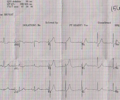
The patient presented with chestpain. If it is STEMI, it would have to be RBBB with STEMI. I was taught that the tell-tale sign of ischemia vs an electrical abnormality was in the hx, i.e. chestpain for the ischemia and potential syncope for brugada. Electrocardiol 45:433-442, 2012 ).
A 70-year-old man calls 911 after experiencing sudden, severe chestpain. The precordial ST-depression pattern on this ECG (and in this clinical setting) should immediately raise suspicion of Posterior STEMI! But if there is none - then you are looking at least at an Isolated Posterior STEMI until proven otherwise.
Edits by Meyers and Smith A man in his 70s with PMH of hypertension, hyperlipidemia, type 2 diabetes, CVA, dual-chamber Medtronic pacemaker, presented to the ED for evaluation of acute chestpain. Code STEMI was activated by the ED physician based on the diagnostic ECG for LAD OMI in ventricular paced rhythm. Limkakeng AT.
A 36 yo male smoker presented to the ED with chestpain. It had started the night before as "indigestion" and had progressed to 8/10 substernal chest pressure radiating to the right shoulder/jaw associated with diaphoresis, nausea, and SOB. of patients with anterior STEMI, ST elevation of greater than or equal to 3.0
A middle aged male presented with chestpain. LVH and the diagnosis of STEMI - how should we apply the current guidelines? In LVH, T-wave inversions are usually much more assymetric , like these (Figure 2): Acute Chestpain, but baseline ECG. How about diagnosing anterior STEMI in the setting of LVH?
This patient presented with a mechanical fall and had chestpain. However, there are also Q-waves inferiorly and the inferior T-waves are inverted, suggesting that this is an old MI with persistent ST elevation, or, alternatively, a subacute or partially reperfused, inferior STEMI. His first troponin I returned at 0.10
He denied chestpain or shortness of breath. In the clinical context of weakness and fever, without chestpain or shortness of breath, the likelihood of Brugada pattern is obviously much higher. Today's patient presented with acute weakness, syncope and fever, but no chestpain or shortness of breath.
2) The STE in V1 and V2 has an R'-wave and downsloping ST segments, very atypical for STEMI. Cardiology was consulted and they agreed that the EKG had an atypical morphology for STEMI and did not activate the cath lab. It was from a patient with chestpain: Note the obvious Brugada pattern. Bicarb 20, Lactate 4.2,
J Electrocardiology 45 (2012):433-442. The patient denied any chestpain whatsoever, and a troponin at zero and 2 hours were both undetectable. Thus, Brugada is the likely diagnosis _ A very nice explanation of this is given in the document quoted below on current ECG criteria for Brugada pattern. Bayes de Luna, A et al.
He had no chestpain, dyspnea, or any other anginal equivalent, and his vital signs were normal. There was indication of parasympathetic overdrive ( the acute inferior STEMI with profound bradycardia and junctional escape ). Multidisciplinary critical care management of electrical storm. link] Mostofsky, E., Maclure, M.,
We organize all of the trending information in your field so you don't have to. Join thousands of users and stay up to date on the latest articles your peers are reading.
You know about us, now we want to get to know you!
Let's personalize your content
Let's get even more personalized
We recognize your account from another site in our network, please click 'Send Email' below to continue with verifying your account and setting a password.


Let's personalize your content