This site uses cookies to improve your experience. To help us insure we adhere to various privacy regulations, please select your country/region of residence. If you do not select a country, we will assume you are from the United States. Select your Cookie Settings or view our Privacy Policy and Terms of Use.
Cookie Settings
Cookies and similar technologies are used on this website for proper function of the website, for tracking performance analytics and for marketing purposes. We and some of our third-party providers may use cookie data for various purposes. Please review the cookie settings below and choose your preference.
Used for the proper function of the website
Used for monitoring website traffic and interactions
Cookie Settings
Cookies and similar technologies are used on this website for proper function of the website, for tracking performance analytics and for marketing purposes. We and some of our third-party providers may use cookie data for various purposes. Please review the cookie settings below and choose your preference.
Strictly Necessary: Used for the proper function of the website
Performance/Analytics: Used for monitoring website traffic and interactions
Whenever a patient does not have chestpain, the pre-test probability of OMI is diminished. Of course SOB, jaw pain, shoulder pain, etc can be a result of OMI, but the pretest probability is less and so you must scrutinize further. Here is the first ED ECG: COMPUTER INTERPRETATION: Electronic Atrial Pacemaker.
The patient presented due to chestpain that was typical in nature, retrosternal and radiating to the left arm and neck. He denied any exertional chestpain. It is unclear if the patient was pain free at this time. He has a medical hx notable for hypertension, hyperlipidemia and previous tobacco use disorder.
He learned more about the patient: A 77 year old female with a past medical history of hypertension and hyperlipidemia presented to the ED at around 0520 after waking up at 0400 with 10/10 chest heaviness radiating to both arms. The patient had continued to have chestpain. He was a paramedic at the time.
A 70-year-old man calls 911 after experiencing sudden, severe chestpain. 2012 Sep;45(5):463-75. Does routine use of the 15-lead ECG improve the diagnosis of acute myocardial infarction in patients with chestpain? This case comes from Sam Ghali ( @EM_RESUS ). Thanks, Sam! O'Gara et al. Circulation. Am J Cardiol.
Written by Willy Frick A man in his 50s with a history of hypertension, dyslipidemia, type 2 diabetes mellitus, and prior inferior OMI status post DES to his proximal RCA 3 years prior presented to the emergency department at around 3 AM complaining of chestpain onset around 9 PM the evening prior. The following ECG was obtained.
A middle aged male presented with chestpain. In LVH, T-wave inversions are usually much more assymetric , like these (Figure 2): Acute Chestpain, but baseline ECG. And sometimes the lateral T-wave is biphasic, with the terminal portion more upright (Figure 3): Acute chestpain. Baseline EKG, no MI.
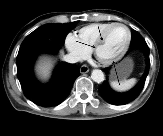
This patient presented with a mechanical fall and had chestpain. His chestpain increased and this ECG was recorded: Now there is increasing inferior ST elevation. Severe Left Main disease, and chestpain with contrast injection into the LM. This case shows a CT image of subendocardial ischemia.
With more than 500 peer-reviewed publications, the HeartFlow FFR CT Analysis remains unparalleled in precision coronary care, as supported by the ACC/AHA ChestPain Guidelines, to improve treatment plans and outcomes. Arbab-Zadeh, Heart Int 2012. 2021 ACC/AHA ChestPain Guidelines. Yokota, et al. Patel et al.
We aimed to examine the diagnostic ability of the score and attempted to improve the predictivity for identifying patients with VSA.Methods:From May 2012 to September 2023, a total 1029 patients underwent ACh provocation test for diagnosing VSA. The original and modified ABCD score were calculated as shown in Figure 1.
The ECG in Figure-1 was obtained from an older woman — who presented with chestpain and palpitations over the previous hour. She had a history of hypertension, and was on medication for this — but she was otherwise healthy. BP = 140/90 mm Hg in association with the rhythm in Figure-1. How would YOU interpret the rhythm in Figure-1 ?
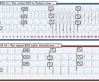
Diagnosis : Atrial flutter with 1:1 conduction, with fast AV conduction made possible by sympathetic drive of exercise On arrival, we obtained another 12-lead: Unremarkable Further history: One month history of shortness of breath on exertion, denies palpitations, chestpain, orthopnea, leg swelling.
She did notice something slightly wrong subjectively, but had no palpitations, chestpain, or SOB, or any other symptom. This middle-aged patient has a remote history of cardiac surgery as a young child for a "heart murmur". Her Apple Watch suddenly told her that she is in atrial fibrillation. She was on no medications.
TheNational Institute for Health and Care Excellence(NICE) recommends CCTA as the first-line investigation for patients with chestpain due to suspected CAD, highlighting its importance in improving diagnostic certainty. 2012) 380:2095128. Eur J Radiol. 2008;66(1):3741. doi:10.1016/j.ejrad.2007.05.006. 2007.05.006. link] iv IMV.2023
The patient presented with chestpain. I was taught that the tell-tale sign of ischemia vs an electrical abnormality was in the hx, i.e. chestpain for the ischemia and potential syncope for brugada. Only 5-18% of ED patients with chestpain have a myocardial infarction of any kind. Is it Brugada pattern?
Edits by Meyers and Smith A man in his 70s with PMH of hypertension, hyperlipidemia, type 2 diabetes, CVA, dual-chamber Medtronic pacemaker, presented to the ED for evaluation of acute chestpain. Annals of Emergency Medicine 2012; 60(12):766-776. Triage ECG: What do you think? This is diagnostic of proximal LAD occlusion.
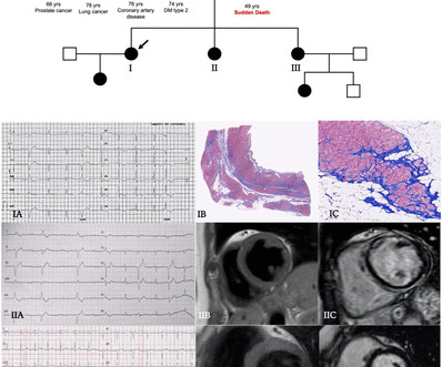
Methods Patients were retrospectively evaluated between January 2012 and June 2020. Clinical contexts leading to diagnosis were SCD in 3 (6%), ventricular arrhythmias in 15 (29%), chestpain in 8 (15%), heart failure in 6 (12%) and familial screening in 20 (38%).
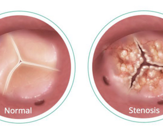
Patients with aortic stenosis often have heart murmurs and experience debilitating symptoms including chestpain, dizziness, fatigue, shortness of breath and an irregular heartbeat. As a result, the heart must work "over time" to pump blood throughout the body.
He denied chestpain or shortness of breath. In the clinical context of weakness and fever, without chestpain or shortness of breath, the likelihood of Brugada pattern is obviously much higher. Today's patient presented with acute weakness, syncope and fever, but no chestpain or shortness of breath.
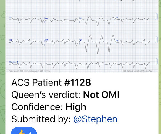
All of the patients presented with chestpain , and they are all in triage. Triage is backed up, and 10 minutes into your shift one of the ED nurses brings your several ECG s that has not been overread by a physician. Which, if any, of these patients has OMI, with myocardium at risk and need for emergent PCI?
Patients with aortic stenosis often have heart murmurs and experience debilitating symptoms including chestpain, dizziness, fatigue, shortness of breath and an irregular heartbeat. As a result, the heart must work "over time" to pump blood throughout the body.
A 36 yo male smoker presented to the ED with chestpain. It had started the night before as "indigestion" and had progressed to 8/10 substernal chest pressure radiating to the right shoulder/jaw associated with diaphoresis, nausea, and SOB. Wong, 2012) STE in aVR of at least 0.5 Wong, 2012) STE in aVR of at least 0.5
She denied chestpain and denied feeling any palpitations, even during her triage ECG: What do you think? e5 Article Download PDF Google Scholar 3 RNW Hauer, MGPJ Cox, JA Groeneweg Impact of new electrocardiographic criteria in arrhythmogenic cardiomyopathy Front Physiol, 3 (2012), p. J Electrocardiol, 42 (2009), pp.
Methods Patients admitted to a single center with acute myocardial infarction (MI) between 1 January 2012 and 31 December 2018, were identified by chart and angiographic review. For secondary outcomes, rates of CVA were 1.7%, chestpain readmission was 22.4%, and repeat angiography was 8.9%. Rates of nonfatal MI were 6.3%
It was from a patient with chestpain: Note the obvious Brugada pattern. The elevated troponin was attributed to either type 2 MI or to non-MI acute myocardial injury. There is no further workup at this time. Smith: Here is a case that was just texted to me today from a former resident. This patient ruled out for MI.
J Electrocardiology 45 (2012):433-442. The patient denied any chestpain whatsoever, and a troponin at zero and 2 hours were both undetectable. Thus, Brugada is the likely diagnosis _ A very nice explanation of this is given in the document quoted below on current ECG criteria for Brugada pattern. Bayes de Luna, A et al.
He had no chestpain, dyspnea, or any other anginal equivalent, and his vital signs were normal. The cardiologist thought she had stent thrombosis which is possible, but I do not necessarily think is sufficient to explain her complete hemodynamic collapse. Multidisciplinary critical care management of electrical storm. Maclure, M.,
We organize all of the trending information in your field so you don't have to. Join thousands of users and stay up to date on the latest articles your peers are reading.
You know about us, now we want to get to know you!
Let's personalize your content
Let's get even more personalized
We recognize your account from another site in our network, please click 'Send Email' below to continue with verifying your account and setting a password.


Let's personalize your content