This site uses cookies to improve your experience. To help us insure we adhere to various privacy regulations, please select your country/region of residence. If you do not select a country, we will assume you are from the United States. Select your Cookie Settings or view our Privacy Policy and Terms of Use.
Cookie Settings
Cookies and similar technologies are used on this website for proper function of the website, for tracking performance analytics and for marketing purposes. We and some of our third-party providers may use cookie data for various purposes. Please review the cookie settings below and choose your preference.
Used for the proper function of the website
Used for monitoring website traffic and interactions
Cookie Settings
Cookies and similar technologies are used on this website for proper function of the website, for tracking performance analytics and for marketing purposes. We and some of our third-party providers may use cookie data for various purposes. Please review the cookie settings below and choose your preference.
Strictly Necessary: Used for the proper function of the website
Performance/Analytics: Used for monitoring website traffic and interactions
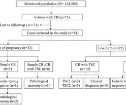
Objective The study aims to assess the ultrasonic features of fetal cardiac rhabdomyoma (CR), track the perinatal outcome and postnatal disease progression, investigate the clinical utility of ultrasound, MRI and tuberous sclerosis complex (TSC) gene analysis in CR evaluation, and offer evidence for determing of fetal CR prognosis.
Annals of Emergency Medicine 2011; Suppl 58(4): S211. And angiographers tell me that it is sometimes difficult to say for certain based on angiogram alone, without intravascular ultrasound or, better yet, optical coherence tomography. Murakami MM. Assuming that was indeed a culprit, then this was ACS.
J Cardiovasc Ultrasound. J Cardiovasc Ultrasound. 2011 Dec;19(4):169-73. Ha J et al. Therapeutic strategies for diastolic dysfunction: a clinical perspective. 2009 Sep;17(3):86-95. Park JH et al. Use and Limitations of E/e’ to Assess Left Ventricular Filling Pressure by Echocardiography. Møller JE et al.
Of interest — the ectopic beats triggering PMVT/VFib in such studies were often mapped to endocardial sites displaying Purkinje potentials within the myocardial scar — suggesting potential responsivity to a 1A agent ( Nogami — Pacing Clin Electrophysiol 34(8): 1034-1049, 2011 ). Administration of Procainamide is 10-17 mg/kg at 20 mg/min.
My bedside ultrasound was of insufficient quality, but showed somewhat reduced overall EF, distended IVC without respiratory variation, no pericardial effusion, and diffuse bilateral B lines. == What do you think of her ECG? J Electrocardiol, 42 (2009), pp.
You use an ultrasound. 2011 Mar-Apr;5(2):105-13. Regardless of the murmur findings they describe. Because using the sounds of a murmur you hear with a stethoscope that was invented in the 1700s is NOT how you make a diagnosis today. Which can now be used easily at the bedside. i.e. You DO NOT GUESS! You measure it DIRECTLY!
Smith comment: This patient did not have a bedside ultrasound. Had one been done, it would have shown a feature that is apparent on this ultrasound (however, this patient's LV function would not be as good as in this clip): This is recorded with the LV on the right. In fact, bedside ultrasound might even find severe aortic stenosis.
We organize all of the trending information in your field so you don't have to. Join thousands of users and stay up to date on the latest articles your peers are reading.
You know about us, now we want to get to know you!
Let's personalize your content
Let's get even more personalized
We recognize your account from another site in our network, please click 'Send Email' below to continue with verifying your account and setting a password.


Let's personalize your content