This site uses cookies to improve your experience. To help us insure we adhere to various privacy regulations, please select your country/region of residence. If you do not select a country, we will assume you are from the United States. Select your Cookie Settings or view our Privacy Policy and Terms of Use.
Cookie Settings
Cookies and similar technologies are used on this website for proper function of the website, for tracking performance analytics and for marketing purposes. We and some of our third-party providers may use cookie data for various purposes. Please review the cookie settings below and choose your preference.
Used for the proper function of the website
Used for monitoring website traffic and interactions
Cookie Settings
Cookies and similar technologies are used on this website for proper function of the website, for tracking performance analytics and for marketing purposes. We and some of our third-party providers may use cookie data for various purposes. Please review the cookie settings below and choose your preference.
Strictly Necessary: Used for the proper function of the website
Performance/Analytics: Used for monitoring website traffic and interactions
The cardiologist recognized that there were EKG changes, but did not take the patient for emergent catheterization because the EKG was “not meeting criteria for STEMI”. Annals of Emergency Medicine 2011; Suppl 58(4): S211. Most STEMI have peak troponin I over 1000 ng/L and most NSTEMI below that level. Murakami MM.
He called 911 and paramedics recorded a prehospital 12 lead ECG which showed a clear inferior STEMI (not shown, tracing could not be found). Research presented at 2011 SAEM in Boston. Objectives : To find the incidence of any rSTD or T-wave inversion (TWI) in angiographically proven inferior STEMI.
Two recent interventions have proven in randomized trials to improve neurologic survival in cardiac arrest: 1) the combination of the ResQPod and the ResQPump (suction device for compression-decompression CPR -- Lancet 2011 ) and 2) Dual Sequential defibrillation. Finally, head-up CPR (which was not used here), makes for better resuscitation.
2011 Dec, 4 (12) 1320–1323 Acquired mimickers of left main atresia 1. We know, how adverse is the outcome of Left main STEMI. If absolutely asymptomatic, and the stress test is negative, leaving it, as it is, is not a forbidden option, in spite of the fact, that the patient would have a single coronary arterial supply. Syphilis 2.
A prehospital ECG was recorded (not shown and not seen by me) which was worrisome for STEMI. A previous ECG from 4 years prior was normal: This looks like an anterior STEMI, but it is complicated by tachycardia (which can greatly elevate ST segments) and by the presentation which is of fever and sepsis.
KEY Point: In areas of the heart where an acute STEMI produces ST elevation — reperfusion T waves ( that develop after the "culprit" artery reopens ) will appear as T wave inversion. In 2011 — Niu et al described the presence of an "N-Wave" — or delayed activation wave of the left ventricular basal region. What is an " N -Wave" ?
Clinical Course The paramedic activated a “Code STEMI” alert and transported the patient nearly 50 miles to the closest tertiary medical center. 2 The astute paramedic recognized this possibility and announced a CODE STEMI. Heart 2011; 97 : 838-843 [link] 14. Look at the aortic outflow tract. What do you see? J Am Coll Cardiol.
Methods This study included consecutive patients with iSTEMI treated with percutaneous coronary intervention (PCI) between 1 January 2011 and 15 July 2019 at a single, tertiary referral centre. Quality initiatives aimed at improving the care of this vulnerable, yet understudied population are needed.
1 in 2011 and 1.73:1 1 in 2011 and 2.11:1 Rates of cardiac catheterization, percutaneous coronary intervention, and coronary artery bypass graft surgery were lower for females than males for STEMI in all countries and years (eg, US cardiac catheterization in 2018, 88.6% 1 in 2018; Israel NSTEMI ratio, 1.71:1 1 in 2018).
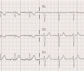
The ECG shows obvious STEMI(+) OMI due to probable proximal LAD occlusion. The patient in today’s case is a previously healthy 40-something male who contacted EMS due to acute onset crushing chest pain. The pain was 10/10 in intensity radiating bilaterally to the shoulders and also to the left arm and neck. The below ECG was recorded.
BP 142/100 HR 90 RR 16 (BBS CTA) SpO2 99 (RA) Dstick 110 My colleagues noted the ST-depression in the respective leads, as well, and STEMI activated to the nearest PCI center. 1] Here is the admitting ED ECG after cancellation of Code STEMI. The EMS crews were correct moving forward with STEMI activation. 1] Driver, B.
A 12-lead was recorded, showing "STEMI," but is unavailable. Moreover, if you know that catastrophic intracranial hemorrhage can result in an ECG that mimics STEMI, then you know that this patient probably has a severe intracranial hemorrhage. She was BVM ventilated and suctioned. Shortly thereafter, pulses were lost.
We organize all of the trending information in your field so you don't have to. Join thousands of users and stay up to date on the latest articles your peers are reading.
You know about us, now we want to get to know you!
Let's personalize your content
Let's get even more personalized
We recognize your account from another site in our network, please click 'Send Email' below to continue with verifying your account and setting a password.


Let's personalize your content