This site uses cookies to improve your experience. To help us insure we adhere to various privacy regulations, please select your country/region of residence. If you do not select a country, we will assume you are from the United States. Select your Cookie Settings or view our Privacy Policy and Terms of Use.
Cookie Settings
Cookies and similar technologies are used on this website for proper function of the website, for tracking performance analytics and for marketing purposes. We and some of our third-party providers may use cookie data for various purposes. Please review the cookie settings below and choose your preference.
Used for the proper function of the website
Used for monitoring website traffic and interactions
Cookie Settings
Cookies and similar technologies are used on this website for proper function of the website, for tracking performance analytics and for marketing purposes. We and some of our third-party providers may use cookie data for various purposes. Please review the cookie settings below and choose your preference.
Strictly Necessary: Used for the proper function of the website
Performance/Analytics: Used for monitoring website traffic and interactions
Cohorts were grouped into preallocation and postallocation change eras: pediatric patients from January 1, 2011, to March 21, 2016, and January 1, 2017, to December 31, 2021; and adult patients from January 1, 2015, to October 17, 2018, and January 1, 2019, to December 31, 2021.
BackgroundCurrent guidelines recommend revascularization in patients with ischemic cardiomyopathy (ICM). A decline in CABG procedures was observed from 2007 to 2011 (annual percentage change, −11.5%;P=0.003), P=0.003), followed by stabilization. CABG and PCI had comparable 30‐day risk‐adjusted mortality risks.
Abstract Introduction It remains unclear if pacing induced cardiomyopathy (PICM) may be minimized by standard pacing of the right ventricle (RV) at sites other than the RV apex. Methods and Results A retrospective evaluation was performed on all patients undergoing pacemaker implantation between 2011 and 2022. PICM occurred in 4.5%
Overall CMR findings are consistent with arrhythmogenic cardiomyopathy. Here is a 2017 review article on ARVD in the New England Journal There is a 2010 publication by the Task Force in Diagnosis of ARVD: Diagnosis of arrhythmogenic right ventricular cardiomyopathy/dysplasia: proposed modification of the task force criteria.
Chagas disease (ChD), prevalent in Brazil, is associated with increased ventricular tachycardia (VT) and ventricular fibrillation (VF) events and SCD compared to other cardiomyopathies. Time periods were chosen based on the establishment of the Arrhythmia Service in 2011. Results Of the 885 patients included, 31% had ChD.
In addition — there seemed to be significant fragmentation ( excessive notching of the QRS ) — which usually is indication of underlying "scar" from infarction, cardiomyopathy, or other form of underlying structural disease. The patient's younger brother was also diagnosed with "a cardiomyopathy". MRI confirmed ARVC.
Director of the Hypertrophic Cardiomyopathy Center at the Lahey Hospital and Medical Center. Impact of Aficamten on Disease and Symptom Burden in Obstructive Hypertrophic Cardiomyopathy: Results from SEQUOIA-HCM. Occurrence of Clinically Diagnosed Hypertrophic Cardiomyopathy in the United States. 30, 2024 — Cytokinetics, Inc.
2017 ) Clinical implication of such coronary anomalies Apart from angiographic surprises, these anomalous coronary arteries may under-perfuse the ventricle and present as unexplained cardiomyopathy , until we realize the anatomical errors in coronary anatomy. 2011 Dec, 4 (12) 1320–1323 Acquired mimickers of left main atresia 1.
Brandão M, Desmoplakin Cardiomyopathy: Comprehensive Review of an Increasingly Recognized Entity. The 2020 “Padua Criteria” for Diagnosis and Phenotype Characterization of Arrhythmogenic Cardiomyopathy in Clinical Practice. J Clin Med. Circulation. 1978 Aug;58(2):305-14. doi: 10.1161/01.cir.58.2.305. PMID: 668079.
A triphasic left ventricular filling pattern with an additional mid diastolic wave, called T wave by some authors and L wave by others, can occur in situations of left ventricular diastolic dysfunction, especially in hypertrophic cardiomyopathy. 2011 Dec;19(4):169-73. J Cardiovasc Ultrasound. 2009 Sep;17(3):86-95. Park JH et al.
The aim of this study was to assess exercise intolerance in FD and identify whether this correlates with the phase of cardiomyopathy. Methods This was a retrospective observational study of adults with FD undergoing cardiopulmonary exercise testing (CPEX) between September 2011 and September 2023 at a national referral centre in the UK.
In most cases, rather, the culprit is gross ischemia due to myocardial infarction, cardiomyopathy, or advanced coronary artery disease. Unfortunately, today’s case is lacking any such diagnostics, thus I cannot say with certainty that the QT interval is, or is not, culpable in arrhythmogenesis. [1] Elsevier: Philadelphia, PA. [7] 8] Liu, E.,
In this abstract from 2011, we found that 4%(4 of 99) type 2 MI and 38% of type 1 MI had ST Elevation. I said I think there is a fixed stenosis in the LAD and the tachycardia and stress caused a type 2 STEMI. link] An angiogram was done: It showed no culprit and no coronary disease, but did show a myocardial bridge in the mid LAD.
Institutional Coronary Artery Bypass Case Volumes and Outcomes European Journal of Heart Failure October 2023 Makoto Mori Robotic Mitral Valve Repair for Degenerative Mitral Regurgitation The Annals of Thoracic Surgery August 2023 Carlos Diaz-Castrillion Volume-Failure to Rescue Relationship in Acute Type A Aortic Dissections: An Analysis of The Society (..)
Institutional Coronary Artery Bypass Case Volumes and Outcomes European Journal of Heart Failure October 2023 Makoto Mori 1 Robotic Mitral Valve Repair for Degenerative Mitral Regurgitation The Annals of Thoracic Surgery August 2023 Carlos Diaz-Castrillion 2 Volume-Failure to Rescue Relationship in Acute Type A Aortic Dissections: An Analysis of The (..)
By ECG alone: it is suspicious for stress cardiomyopathy, or takotsubo, due to the diffuse ST Elevation: II, III, aVF AND I and aVL. Subarachnoid hemorrhage causes extreme central catecholamine output, resulting in stress cardiomyopathy, just like takotsubo. This is unusual in acute OMI. she had severe pulmonary edema.
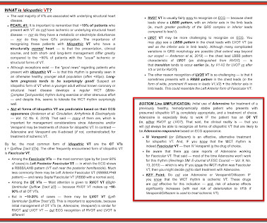
Whenever I see PVCs with the morphology and axis seen in todays case I always look for signs of AC ( Arrhythmogenic Cardiomyopathy ). Arrhythmogenic cardiomyopathy often manifests with PVCs from the RV. The ECG in Figure-1 however, shows no signs of arrhythmogenic cardiomyopathy.
We organize all of the trending information in your field so you don't have to. Join thousands of users and stay up to date on the latest articles your peers are reading.
You know about us, now we want to get to know you!
Let's personalize your content
Let's get even more personalized
We recognize your account from another site in our network, please click 'Send Email' below to continue with verifying your account and setting a password.


Let's personalize your content