This site uses cookies to improve your experience. To help us insure we adhere to various privacy regulations, please select your country/region of residence. If you do not select a country, we will assume you are from the United States. Select your Cookie Settings or view our Privacy Policy and Terms of Use.
Cookie Settings
Cookies and similar technologies are used on this website for proper function of the website, for tracking performance analytics and for marketing purposes. We and some of our third-party providers may use cookie data for various purposes. Please review the cookie settings below and choose your preference.
Used for the proper function of the website
Used for monitoring website traffic and interactions
Cookie Settings
Cookies and similar technologies are used on this website for proper function of the website, for tracking performance analytics and for marketing purposes. We and some of our third-party providers may use cookie data for various purposes. Please review the cookie settings below and choose your preference.
Strictly Necessary: Used for the proper function of the website
Performance/Analytics: Used for monitoring website traffic and interactions
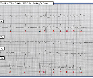
It is atrialflutter with 2:1 conduction. There are clear flutter waves in lead II across the bottom. In V1, there are upright waves that appear to be P-waves but are not: they are atrial waves and it is typical for atrialflutter waves to be upright in V1, whereas sinus P-waves are biphasic in V1.
Two recent interventions have proven in randomized trials to improve neurologic survival in cardiac arrest: 1) the combination of the ResQPod and the ResQPump (suction device for compression-decompression CPR -- Lancet 2011 ) and 2) Dual Sequential defibrillation. Finally, head-up CPR (which was not used here), makes for better resuscitation.
By this definition, a variety of rhythms may qualify as “SVTs” — including sinus tachycardia, atrialflutter or fibrillation, MAT, AVRT/AVNRT, among others. ECG Blog #138 — AFlutter vs Atrial Tachycardia. ECG Blog #40 — Another regular SVT that turned out to be AFlutter.
PEARL # 3: At this point — the most time-efficient step for solving today's rhythm will be to determine the nature of atrial activity. This type of Wenckebach response that may be seen with atrial tachycardia ( or atrialflutter ) — is often physiologic, as a result of the rapid atrial rate that occurs with these arrhythmias.
Smith comment-2: Another adverse effect from flecainide is atrialflutter with 1:1 conduction (if you happen to go into atrialflutter, the flecainide slows the flutter rate such that it is slow enough to conduct throught the AV node at 1:1 and you can end up with a ventricular rate of 220!!
We organize all of the trending information in your field so you don't have to. Join thousands of users and stay up to date on the latest articles your peers are reading.
You know about us, now we want to get to know you!
Let's personalize your content
Let's get even more personalized
We recognize your account from another site in our network, please click 'Send Email' below to continue with verifying your account and setting a password.


Let's personalize your content