This site uses cookies to improve your experience. To help us insure we adhere to various privacy regulations, please select your country/region of residence. If you do not select a country, we will assume you are from the United States. Select your Cookie Settings or view our Privacy Policy and Terms of Use.
Cookie Settings
Cookies and similar technologies are used on this website for proper function of the website, for tracking performance analytics and for marketing purposes. We and some of our third-party providers may use cookie data for various purposes. Please review the cookie settings below and choose your preference.
Used for the proper function of the website
Used for monitoring website traffic and interactions
Cookie Settings
Cookies and similar technologies are used on this website for proper function of the website, for tracking performance analytics and for marketing purposes. We and some of our third-party providers may use cookie data for various purposes. Please review the cookie settings below and choose your preference.
Strictly Necessary: Used for the proper function of the website
Performance/Analytics: Used for monitoring website traffic and interactions
Distribution Variance of Focal Atrial Tachycardia Foci and Long-Term Outcomes After Ablation. ABSTRACT Introduction The distribution of the origin of focal atrial tachycardia (FAT) in patients with different ages have not been clearly elucidated. Conclusion Significant age differences were observed in the distribution of FAT foci.
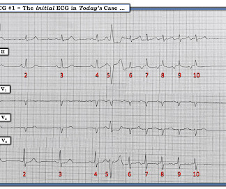
A series of cardiac arrhythmias were seen during the course of her resuscitation — including the interesting arrhythmia shown in the long lead II of Figure-1. PEARL # 5: The simple act of labeling P waves can be invaluable for solving an arrhythmia. Figure-6: Laddergram illustration of the mechanism in today's arrhythmia.
This may explain the poorer response of pleomorphic VT to antiarrhythmic therapy — and the higher morbidity and mortality that seems to be associated with this arrhythmia. This entity appears to predispose to unstable reentrant conditions that increase the chance of deterioration from VT to PMVT or VFib.
By this definition, a variety of rhythms may qualify as “SVTs” — including sinus tachycardia, atrial flutter or fibrillation, MAT, AVRT/AVNRT, among others. Why Isn’t this a Run of Atrial Tachycardia? — ECG Blog #185 — Reviews the P s, Q s, 3 R Approach to Arrhythmia Interpretation.
myocardial infarction), arrhythmias, valvular pathology, shunts, or outflow obstructions. Heart 2011; 97 : 838-843 [link] 14. Hypotension may of course be a result of a brady- or tachydysrhythmia. 2) Hypoxia, including poisons of oxidative phosphorylation such as HS, CO, CN. J Am Coll Cardiol. 1985;5(3):711-716.
She was awake, alert, well perfused, with normal mental status and overall unremarkable physical exam except for a regular tachycardia, possible rales at both bases, some mild RUQ abdominal tenderness. Thus, I believe it is a regular, monomorphic, wide complex tachycardia. Or it could simply still be classic VT. What is the Diagnosis?
What is the most likely cause of this arrhythmia? IMPRESSION: Given the presence of a wide tachycardia — with 2 distinct QRS morphologies, and no sign of P waves — a presumed diagnosis of B i D irectional Ventricular Tachycardia has to be made. Multifocal vs Polymorphic VT — September 23, 2011 post from Dr. S.
However, he suddenly developed a series of malignant ventricular arrhythmias. This progressed to electrical storm , with incessant PolyMorphic Ventricular Tachycardia ( PMVT ) and recurrent episodes of Ventricular Fibrillation ( VFib ). Below are printouts of some of the arrhythmias recorded. What do you think?
We aimed to investigate the prevalence of NSVT in patients with ATTRwr-CA, and the association of NSVT with sustained ventricular arrhythmias (VA) and all-cause mortality.
Chagas disease (ChD) was associated with increased rates of ventricular tachycardia and ventricular fibrillation in ICD patients only in the initial two periods, but there was no statistical difference in the last period. Time periods were chosen based on the establishment of the Arrhythmia Service in 2011.
During observation in the ED the patient had multiple self-terminating runs of Non-Sustained monomorphic Ventricular Tachycardia (NSVT). The possibility of an ischemic cause of the ventricular arrhythmia has to be considered! A workup was undertaken in search of a cause of the patient's ventricular arrhythmia. No PVCs are seen.
We organize all of the trending information in your field so you don't have to. Join thousands of users and stay up to date on the latest articles your peers are reading.
You know about us, now we want to get to know you!
Let's personalize your content
Let's get even more personalized
We recognize your account from another site in our network, please click 'Send Email' below to continue with verifying your account and setting a password.


Let's personalize your content