This site uses cookies to improve your experience. To help us insure we adhere to various privacy regulations, please select your country/region of residence. If you do not select a country, we will assume you are from the United States. Select your Cookie Settings or view our Privacy Policy and Terms of Use.
Cookie Settings
Cookies and similar technologies are used on this website for proper function of the website, for tracking performance analytics and for marketing purposes. We and some of our third-party providers may use cookie data for various purposes. Please review the cookie settings below and choose your preference.
Used for the proper function of the website
Used for monitoring website traffic and interactions
Cookie Settings
Cookies and similar technologies are used on this website for proper function of the website, for tracking performance analytics and for marketing purposes. We and some of our third-party providers may use cookie data for various purposes. Please review the cookie settings below and choose your preference.
Strictly Necessary: Used for the proper function of the website
Performance/Analytics: Used for monitoring website traffic and interactions
Methods and Results A total of 323 consecutive FAT patients who underwent electrophysiological study and radiofrequency catheter ablation between January 2011 and March 2023 were selected for this study. Conclusion Significant age differences were observed in the distribution of FAT foci.
However, he suddenly developed a series of malignant ventricular arrhythmias. Below are printouts of some of the arrhythmias recorded. This time, the arrhythmia did not spontaneously terminate — but rather degenerated to VFib, requiring defibrillation. The arrhythmia starts with a PVC having a short coupling interval.
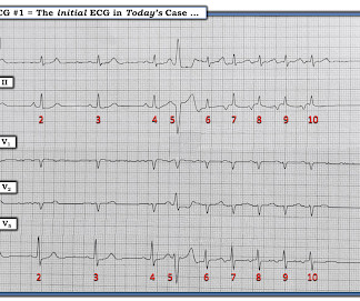
What is the most likely cause of this arrhythmia? Multifocal vs Polymorphic VT — September 23, 2011 post from Dr. S. He developed cardiac arrest shortly after the ECG in Figure-1 was recorded. QUESTIONS: How would YOU interpret the ECG in Figure-1 ? Figure-1: The initial ECG in today's case. (
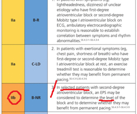
Massie Block -Ref 1) But, if there is something unusual in the clinical history, be ready to investigate until the arrhythmia, or at least the anxiety disappears. Final message AV blocks, even Mobitz type 2, can occur at normal times of heightened vagal tone.( Reference 1. Circulation. 1978 Aug;58(2):305-14. doi: 10.1161/01.cir.58.2.305.
Methods and Results All consecutive patients ( n =264) with previous ICD who underwent LVAD surgery between May 2011 and December 2019 at our institution were included. ABSTRACT Aim To evaluate the predictive value of preoperative echocardiographic parameters for occurrence of VAs in patients with preexisting ICD undergoing LVAD implantation.
The study aims to assess whether mortality and VT/VF events decreased in patients who received ICDs during different time periods (2007–2010, 2011–2014, and 2015–2018). Time periods were chosen based on the establishment of the Arrhythmia Service in 2011. 001) and the combined outcome ( p = .009).
Consecutive patients admitted to coronary care unit (CCU) were enrolled between January 2011 and December 2020. Among them, 1,129 (28.2%) had acute coronary syndrome, 1,915 (47.8%) had coronary artery disease, 1,039 (25.9%) had arrhythmia, and 1,825 (45.6%) had heart failure.
We aimed to investigate the prevalence of NSVT in patients with ATTRwr-CA, and the association of NSVT with sustained ventricular arrhythmias (VA) and all-cause mortality. Patients with NSVT have a higher risk of incident sustained VT compared to those without NSVT.
Free full text: [link] There are 6 categories of criteria : 1) Imaging 2) Pathologic 3) ECG Repolarization 4) ECG Depolarization 5) Arrhythmias 6) Family History. J Electrocardiol, 42 (2009), pp. Of Note: This patient was hemodynamically stable without palpitations at the time ECG #1 was recorded.
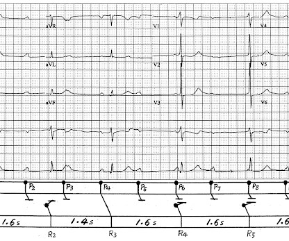
A series of cardiac arrhythmias were seen during the course of her resuscitation — including the interesting arrhythmia shown in the long lead II of Figure-1. PEARL # 5: The simple act of labeling P waves can be invaluable for solving an arrhythmia. Figure-6: Laddergram illustration of the mechanism in today's arrhythmia.
A good example of the same situation was posted at “theheart.org” arrhythmia/EP section “ECG of the month” program in 2/2012. Here is another case posted in June 2011. AV junctional escape and AV dissociation are all obligatory secondary responses.
This is a circumstance in which there exists a single focus of arrhythmogensis, yet conducts through multiple exit sites and/or experiences shifting conduction properties for arrhythmia duration. Chapter 17: Ventricular Arrhythmias (pg. Chapter 17: Ventricular Arrhythmias (pg. Chapter 16: Ventricular Arrhythmias (pg.
ECG Blog #185 — Reviews the P s, Q s, 3 R Approach to Arrhythmia Interpretation. González-Torrecilla et al: Ann Noninvasive Electrocardiol 16(1):85-95, 2011 — Reviews distinction between AVNRT vs AVRT and other regular SVT rhythms in patients without WPW. ECG Blog #138 — AFlutter vs Atrial Tachycardia.
This may explain the poorer response of pleomorphic VT to antiarrhythmic therapy — and the higher morbidity and mortality that seems to be associated with this arrhythmia. This entity appears to predispose to unstable reentrant conditions that increase the chance of deterioration from VT to PMVT or VFib.
myocardial infarction), arrhythmias, valvular pathology, shunts, or outflow obstructions. Heart 2011; 97 : 838-843 [link] 14. Fundamentally, cardiogenic shock is an issue of decreased cardiac output. This may be secondary to multiple factors, including decreased cardiac contractility (ie. J Am Coll Cardiol. 1985;5(3):711-716.
My Thoughts on the ECG in Figure-1: The rhythm in ECG #1 is sinus arrhythmia. Figure-1: Potential Causes of acute MI in Children ( Adapted from Suryawanshi et al — Ann Pediatr Cardiol 4(1):81-83, 2011 ). Figure-1: I've labeled the initial ECG in today's case. As per Drs.
The possibility of an ischemic cause of the ventricular arrhythmia has to be considered! A workup was undertaken in search of a cause of the patient's ventricular arrhythmia. Once the arrhythmia was under control cardiac MRi was performed. The idiopathic VTs are an interesting group of arrhythmias! No PVCs are seen.
We organize all of the trending information in your field so you don't have to. Join thousands of users and stay up to date on the latest articles your peers are reading.
You know about us, now we want to get to know you!
Let's personalize your content
Let's get even more personalized
We recognize your account from another site in our network, please click 'Send Email' below to continue with verifying your account and setting a password.


Let's personalize your content