This site uses cookies to improve your experience. To help us insure we adhere to various privacy regulations, please select your country/region of residence. If you do not select a country, we will assume you are from the United States. Select your Cookie Settings or view our Privacy Policy and Terms of Use.
Cookie Settings
Cookies and similar technologies are used on this website for proper function of the website, for tracking performance analytics and for marketing purposes. We and some of our third-party providers may use cookie data for various purposes. Please review the cookie settings below and choose your preference.
Used for the proper function of the website
Used for monitoring website traffic and interactions
Cookie Settings
Cookies and similar technologies are used on this website for proper function of the website, for tracking performance analytics and for marketing purposes. We and some of our third-party providers may use cookie data for various purposes. Please review the cookie settings below and choose your preference.
Strictly Necessary: Used for the proper function of the website
Performance/Analytics: Used for monitoring website traffic and interactions
She was awake, alert, well perfused, with normal mental status and overall unremarkable physical exam except for a regular tachycardia, possible rales at both bases, some mild RUQ abdominal tenderness. Thus, I believe it is a regular, monomorphic, wide complex tachycardia. Or it could simply still be classic VT. What is the Diagnosis?
Abstract Introduction Supraventricular tachycardia (SVT) is a common pediatric arrhythmia. Individuals aged 1–21 years at time of SVT diagnosis initiated on a BB or a CCB between 01/01/2010 and 12/31/2020 were included. Exclusion criteria were pre-excitation, ectopic atrial tachycardia, and hemodynamically significant heart disease.
They had already cardioverted at 120 J, then 200 J, which resulted in the following: Ventricular Tachycardia They then cardioverted at 200 J which r esulted in the same narrow complex rhythm shown above, at 185 beats per minute. This would treat both SVT or sinus tachycardia. I suggested esmolol if the heart rate did not improve.
Given the efficacy of other class III agents, it has been used off-label for the treatment of premature ventricular complexes (PVCs) and ventricular tachycardias (VTs). Among the 32 patients with PVCs who successfully started dofetilide, the mean PVC burden decreased from 2010% to 88% at a median follow-up of 2.6 months ( p <.001).
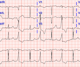
KEY Point: Look for additional simultaneously-recorded leads = “12 Leads are Better than One!” ( ie, For example with tachycardias — the QRS may look narrow if all you have is 1 or 2 leads — whereas if part of the QRS lies on the baseline in the single lead you are looking at, this might be VT! ). 19:50 — Not appreciating statistical odds! (
Chagas disease (ChD) was associated with increased rates of ventricular tachycardia and ventricular fibrillation in ICD patients only in the initial two periods, but there was no statistical difference in the last period. Progressive decline across periods in mortality rates among patients with implantable cardioverter-defibrillator (ICD).
Another way that WPW can be concealed is in the very rare (~15% of all WPW patients) retrograde-only conduction, in which the accessory pathway ONLY allows retrograde conduction, which obviously wouldn't show a delta wave on sinus EKG but still predisposes the patient to re-entry tachycardias. 2010 Mar-Apr;43(2):144-5. References: 1.
22:25 — What if you have a regular SVT ( = narrow-complex tachycardia ) without obvious P waves? ( 12:15 — Regarding my experience from the 1980s until ~2010: How I went from hating computer interpretations to loving them ( after I finally understood what the computer can and can not do ). The 4 common causes? —
NOTE: As discussed in detail in ECG Blog #108 — " A IVR" is an "enhanced" ventricular ectopic rhythm that occurs faster than the intrinsic ventricular escape rate ( which is typically between 20-40/minute ) — but slower than hemodynamically significant Ventricular Tachycardia ( ie, VT at rates >130-140/minute ).
T-wave inversions and dynamic ST elevation Tachycardia, hyperthyroid, and ST elevation. A nice Review of EIA by Molis and Molis can be found in Sports Health 2:311-317, 2010. Hypertrophic Cardiomyopathy or Normal ("Variant")? Two cases of ST Elevation with Terminal T-wave Inversion - do either, neither, or both need reperfusion?
Answer : you must treat the patient's underlying condition causing sinus tachycardia, and repeat the ECG at the lower heart rate. JACC 55(9):934-947; 2010 ]. Optimal QT interval correction formula in sinus tachycardia for identifying cardiovacular and mortality risk: Findings from the Penn Atrial Fibrillation Free study.
By far, the most common etiology of acute pericarditis in a young adult is idiopathic ( Khandaker et al: Mayo Clin Proc 85(6): 572-593, 2010 ) although many ( most ) of these idiopathic cases are probably viral. One looks for sinus tachycardia and diffuse low voltage but many conditions produce these nonspecific findings.
If the patient has Abnormal Vital Signs (fever, hypotension, tachycardia, or tachypnea, or hypoxemia), then these are the primary issue to address, as there is ongoing pathology which must be identified. J Am Coll Cardiol, 2010; 55:713-721, doi:10.1016/j.jacc.2009.09.049 The tracings were considered abnormal in the following cases: 1.
Heart Rhythm 2010 Hudzik B, Gasior M. The relationship between J wave and ventricular tachycardia during Takotsubo cardiomyopathy. Indian Pacing Electrophysiol J 2004 Antzelevitch C, Yan G. J wave syndromes. J-waves in hypothermia. CMAJ 2017 Vassallo SU, Delaney KA, Hoffman RS, et al.
We organize all of the trending information in your field so you don't have to. Join thousands of users and stay up to date on the latest articles your peers are reading.
You know about us, now we want to get to know you!
Let's personalize your content
Let's get even more personalized
We recognize your account from another site in our network, please click 'Send Email' below to continue with verifying your account and setting a password.


Let's personalize your content