This site uses cookies to improve your experience. To help us insure we adhere to various privacy regulations, please select your country/region of residence. If you do not select a country, we will assume you are from the United States. Select your Cookie Settings or view our Privacy Policy and Terms of Use.
Cookie Settings
Cookies and similar technologies are used on this website for proper function of the website, for tracking performance analytics and for marketing purposes. We and some of our third-party providers may use cookie data for various purposes. Please review the cookie settings below and choose your preference.
Used for the proper function of the website
Used for monitoring website traffic and interactions
Cookie Settings
Cookies and similar technologies are used on this website for proper function of the website, for tracking performance analytics and for marketing purposes. We and some of our third-party providers may use cookie data for various purposes. Please review the cookie settings below and choose your preference.
Strictly Necessary: Used for the proper function of the website
Performance/Analytics: Used for monitoring website traffic and interactions
As discussed in ECG Blog #108 — AIVR generally occurs in one of the following C linical S ettings : i ) As a rhythm during cardiacarrest; ii ) In the monitoring phase of acute MI ( especially with inferior MI ) ; or , iii ) As a reperfusion arrhythmia ( ie, following thrombolysis, acute angioplasty, or spontaneous reperfusion ).
The ECG in Figure-1 was obtained from an 18-year old woman — who moments before been resuscitated from out-of-hospital cardiacarrest. Does this ECG in Figure-1 provide clue(s) to the etiology of this patient's cardiacarrest? I suspected the answer resides in the reason why an 18-year woman might have a cardiacarrest.
What is the utility of a head CT in cardiacarrest? We studied this and published the abstract below in 2010. We found intracranial hemorrhage in 2% of non-traumatic cardiacarrest patients, and in 4 others the presence of cerebral edema changed management. Chicago November 2010. Kurkciyan et al.
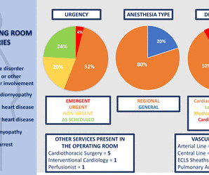
Study design We performed a retrospective evaluation of pregnant patients with high-risk CVD who delivered in the main OR at a large academic centre between January 2010 and March 2021. Results Of 25 deliveries, connective tissue disease (n=9, 36%) was the most common CVD type, followed by non-ischaemic cardiomyopathy (n=5, 20%).
Heart Rhythm 2010 Hudzik B, Gasior M. The relationship between J wave and ventricular tachycardia during Takotsubo cardiomyopathy. The final letter in the SLOWED mnemonic is " D " for "Dead" ( resulting from VT/VF or asystolic cardiacarrest ). Indian Pacing Electrophysiol J 2004 Antzelevitch C, Yan G. J wave syndromes.
We organize all of the trending information in your field so you don't have to. Join thousands of users and stay up to date on the latest articles your peers are reading.
You know about us, now we want to get to know you!
Let's personalize your content
Let's get even more personalized
We recognize your account from another site in our network, please click 'Send Email' below to continue with verifying your account and setting a password.


Let's personalize your content