This site uses cookies to improve your experience. To help us insure we adhere to various privacy regulations, please select your country/region of residence. If you do not select a country, we will assume you are from the United States. Select your Cookie Settings or view our Privacy Policy and Terms of Use.
Cookie Settings
Cookies and similar technologies are used on this website for proper function of the website, for tracking performance analytics and for marketing purposes. We and some of our third-party providers may use cookie data for various purposes. Please review the cookie settings below and choose your preference.
Used for the proper function of the website
Used for monitoring website traffic and interactions
Cookie Settings
Cookies and similar technologies are used on this website for proper function of the website, for tracking performance analytics and for marketing purposes. We and some of our third-party providers may use cookie data for various purposes. Please review the cookie settings below and choose your preference.
Strictly Necessary: Used for the proper function of the website
Performance/Analytics: Used for monitoring website traffic and interactions
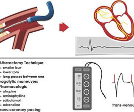
Background Rotational atherectomy (RA) during percutaneous coronary intervention may cause transient bradycardia or a higher-degree heart block. Traditionally, some operators use prophylactic transvenous pacing wire (TPW) to avoid haemodynamic complications associated with bradycardia.
Methods We retrospectively screened 2009 patients who underwent pacemaker implantation from 2010 to 2020 in seven institutions. A CLBBB-like paced QRS was defined as meeting the CLBBB criteria of the American Heart Association/American College of Cardiology Foundation/Heart Rhythm Society in 2009.
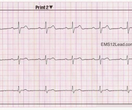
Original publication date July, 2009. We have borderline sinus bradycardia with 1 ° AVB and occasional PACs. ECG diagnosis: Borderline sinus bradycardia, 1st degree AVB, RBBB, and occasional PACs. Concept Review How do you identify right bundle branch block (RBBB) on the 12 lead ECG? What’s the rhythm?
Use of drugs producing bradycardia like beta blockers in stages III and IV may precipitate low output state. 2009 Sep;17(3):86-95. In stage IV, this restrictive filling pattern remains fixed even during Valsalva maneuver. Initial stages (I to III) are considered reversible with treatment. Stage IV is considered as advanced. Ha J et al.
There’s sinus bradycardia, normal conduction, normal axis, delayed R wave progression, and normal voltages. The patient has a history of CABG so some of these changes could be old, but with ongoing chest pain and bradycardia in a high risk patient this is still acute OMI until proven otherwise. Sinus bradycardia.” Busk et al.
PVCs N ot generally considered abnormal ECG findings: Isolated PAC, First Degree AV Block, Sinus bradycardia at a rate of 35-45, and Nonspecific ST-T abnormalities (even if different from a previous ECG). Thus, if there is documented sinus bradycardia, and no suspicion of high grade AV block, at the time of the syncope, this is very useful.
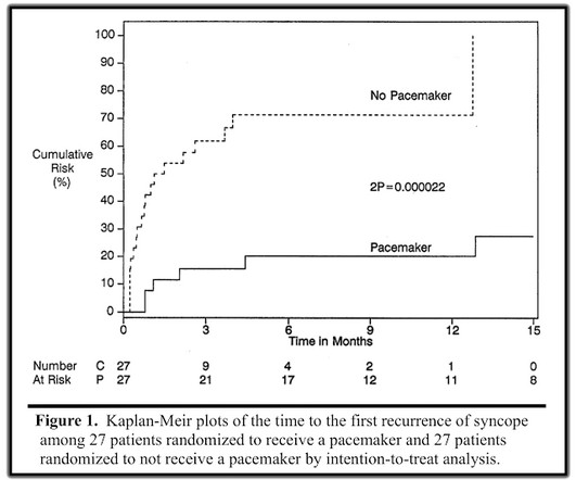
Perhaps because the bradycardia in vasovagal syncope is only one part of the autonomic response. Phase 4 block is also referred to as "bradycardia dependent block." A stunning result. One of many examples in medical history that remind us of the importance of blinding in clinical trials. Why was there no benefit? link] Connolly, S.,
We organize all of the trending information in your field so you don't have to. Join thousands of users and stay up to date on the latest articles your peers are reading.
You know about us, now we want to get to know you!
Let's personalize your content
Let's get even more personalized
We recognize your account from another site in our network, please click 'Send Email' below to continue with verifying your account and setting a password.


Let's personalize your content